Adding intravenous contrast enhancement to virtual colonoscopy has been shown to increase lesion conspicuity, help differentiate polyp from stool, and increase reader confidence. But enhancement does not reliably increase with either polyp size or cancer stage.
In a study published in Radiology Online, researchers from Beth Israel Deaconess Medical Center in Boston found that the degree of enhancement correlated poorly with the degree of differentiation, although grade 1 adenomas did enhance less than lesions grade 2 and higher (Radiology, May 29, 2003).
"The significance of polyp enhancement must be further elucidated," wrote Dr. Jacob Sosna, Dr. Martina Morrin, Dr. Jonathan Kruskal and colleagues. "There is evidence that neovascularization is an early critical event in primary colorectal tumorigenesis, reaching a maximum level early in the malignant process. Vascular branching counts, an indirect estimate of tumor angiogenic activity, are significantly higher in carcinomas than they are in adenomas. For colonic tumors, microvascular density, a direct measurement of angiogenic vessels in a solid tumor, is lower in the normal mucosa than in adenomas, and is highest in carcinomas."
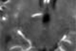
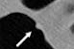
In the study, the researchers retrospectively quantified the degree of contrast enhancement in 29 consecutive neoplasms, both pre- and post-contrast CT, that were subsequently resected. The differences in attenuation pre- and post-contrast were then compared.
"The neoplasms were graded as follows: grade 1, adenoma; grade 2, adenoma with high-grade dysplasia; grade 3, well-differentiated adenocarcinoma; grade 4, moderately differentiated adenocarcinoma; and grade 5, poorly differentiated adenocarcinoma," the authors wrote.
The 29 patients (from a cohort of 642) had undergone contrast-enhanced virtual colonoscopy between 1997 and 2002, following standard bowel preparation one to two days before the procedure with either a standard barium enema preparation (Fleet Prep Kit 1, Fleet Pharmaceutical, Lynchburg, VA) or balanced polyethylene glycol solution (GoLytely, Braintree Laboratories, Braintree, MA).
All patients were scanned in both prone and supine positions using a HiSpeed or LightSpeed QX/i scanner (GE Medical Systems, Waukesha, WI) at 3-mm collimation, pitch of 2, 6-mm/sec table speed, 120 mAs, 120 kVp, and a 512 x 512 matrix. The images were reconstructed at 1.5-mm intervals with a 1.5-mm overlap, and interpreted on a GE Advantage Windows workstation by two experienced radiologists, who resolved interpretive differences by consensus.
"Enhancement values for each lesion were measured by calculating the mean of three attenuation values ... of at least a 1-cm2 ... region of interest obtained before and after contrast enhancement," the authors wrote. "The net change was then calculated as the enhancement." Data analysis used Pearson and Spearman correlation coefficients to assess the correlation between the degree of polyp enhancement and the histologic grade.
According to the results, the neoplasms ranged in size from 10 to 65 mm, with a mean diameter of 27.9 mm. Twelve lesions were grade 1 (59 ± 18 HU), three were grade 2 (49 ± 17 HU), two were grade 3 (56 ± 16 HU), nine were grade 4 (66 ± 22 HU) and two were grade 5 (85 ± 30 HU).
"There was no correlation between size and degree of enhancement, size and histologic grade (R=-0.17, P=33), or histologic grade and degree of enhancement (R=0.23, P=0.23)," Sosna and colleagues wrote. "However, increasing enhancement was noted between grades 2 and 5. When an enhancement threshold of 40 HU was used for the diagnosis of adenocarcinoma (grades 3-5), sensitivity was 92%, specificity was 20%, positive predictive value was 50%, and negative predictive value was 75%."
The lack of correlation between enhancement and either lesion size or grade of malignant differentiation was somewhat surprising, according to the authors, since larger lesions might be expected to show greater enhancement and less differentiation.
"However, tumor perfusion is variable and unpredictable...," they wrote. "Although there seems to be a trend of increasing enhancement from adenoma with dysplasia (grade 2) into poorly differentiated adenocarcinoma (grade 5), it was not significant." Thus, their correlative hypothesis needs to be rejected, the authors concluded.
"We suggest that further studies be performed, not only to increase the number of lesions being analyzed and compared, but also to optimize perfusion parameters for the in vivo characterization of tumor neovascularity," they wrote.
By Eric BarnesAuntMinnie.com staff writer
June 9, 2003
Related Reading
Virtual colonoscopy: to contrast, or not to contrast, April 10, 2002
Copyright © 2003 AuntMinnie.com