Conforming CT protocols to the patient's body mass can help determine the lowest radiation dose needed to acquire diagnostic-quality images. But such adjustments are often made informally if at all, with the radiologist taking a quick look at the patient's weight and/or body habitus before imaging begins.
Now, virtual colonoscopy providers in the Netherlands have found a way to quantify the process without overcomplicating it. At the 2002 RSNA meeting in Chicago, they also sought to quantify the validity of their results.
"The purpose of our study was to maintain constant image quality in CT colonography independent of patient size," said medical physicist Henk Venema, Ph.D., from the Academic Medical Center of the Netherlands in Amsterdam. "The solution, of course, is to tailor the mAs value to patient size; that is, lower mAs for smaller patients, and higher mAs for the thicker ones."
In the study, 50 consecutive patients underwent virtual colonoscopy at a fixed exposure (120 kV, 100 mAs per slice, 4 x 2.5-mm collimation on an Mx8000 multislice CT scanner (Philips Medical Systems, Best, the Netherlands).
Venema and his colleagues, Dr. Rogier van Gelder, Dr. Rogier Determann, Dr. Johan Laméris, and Dr. Jaap Stoker, measured each patient's waist size midway between the lower rib and the lateral iliac crest. To measure overall image quality, they measured the standard deviation (SD) of the image noise at the colon surface in all patients.
"As everyone knows, noise can be easily measured in a homogenous region of interest," he said. "That's a very good indicator of the detectability of small polyps.... And the solution is to measure noise in each pixel in a cross section according to a simple formula."
Waist sizes among the 50 patients ranged from 72 to 118 cm (28.3 to 46.5 inches), mean 98 cm (38.6 inches), SD 0.8. To determine the optimal mAs value for different patient sizes, they divided the patients into three waist-size groups:
- Small: < 87.5 cm (< 34.5 inches).
- Medium: 87.5 to 102.5 cm (34.5 to 40.4 inches).
- Large: > 102.5 cm (> 40.4 inches).
Testing the method in six patients, the estimated and measured SD values in the bladder and aorta were found to be in good agreement, Venema said. The group scaled the noise factors to the mAs values, which resulted in constant image quality for all three groups.
"The frequency distributions of the SD values ranged from 0.6 to more than 3, the mean value (was) 1.2. And they are expressed in a relative scale, relative to the standard patient," he said.
 |
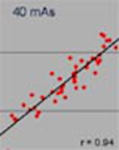
| The charts above show image quality (quantified with the mean standard deviation (SD) at the colon surface) as a function of waist circumference. Higher SD values correspond with lower image quality. The logarithm of the SD is plotted above. The SD values at the left are for a constant mAs value (40 mAs); those at the right for mAs value according to size: 23 mAs for small patients (waist < 87.5 cm or < 35.4 inches); 40 mAs for medium-sized patients (waist 87.5 to 102.5 cm, or 34.5 to 40.4 inches) and 69 mAs for large patients (waist > 102.5 cm or > 40.4 inches). |
 |
| At left, the mean SD values at the colon surface when mAs values are tailored to each waist size individually. The actual mAs values for each waist size are shown at right (from 16 mAs for the smallest patient to 93 mAs for the largest patient, with approximately the same image quality). For comparison purposes, the mAs values used in the first image above are also shown at right. The variation of the SD values with the individual approach is smaller. However, using three size classes is often more convenient in clinical practice. Graphics courtesy of Dr. Henk Venema. |
The aim is to perform virtual colonoscopy with the lowest radiation dose compatible with the detection of clinically relevant polyps, Venema concluded.
"Waist surface as a measure of surface size is, of course, very easy to measure, and you can see that it is a very accurate predictor of the noise value in CT colonography," he said. "In our (virtual colonoscopy) protocol at AMC hospital, we now tailor the mAs to patient size. As a result of the study we have also lowered the mAs values from a standard of 100 mAs to 25 mAs for small patients, 35 mAs for medium patients, and 70 mAs for large patients."
By Eric BarnesAuntMinnie.com staff writer
February 7, 2003
Related Reading
Workflow issues key in virtual colonoscopy, January 15, 2003
Worrisome radiation dose seen in CT lung screening, follow-up, December 23, 2002
No compromise in breast image quality with lower FFDM dose, December 13, 2002
Low-dose CT colonography accurately detects large colorectal neoplasms, July 30, 2002
Copyright © 2003 AuntMinnie.com