CHICAGO - Radiologists and radiographers were nearly equally accurate in evaluating precancerous lesions in patients undergoing virtual colonoscopy, according to a Dutch study presented at the RSNA conference.
Overall, specificity and sensitivity in reading the CT data from 138 patients by radiologists and radiographers were similar, said Dr. Marloes Thomassen-de Graaf, a researcher at the Academic Medical Center in Amsterdam.
“CT colonoscopy evaluation by radiologists may facilitate implementation of this technique in colorectal cancer screening,” she said in her presentation at the 88th annual meeting.
In the study, two radiographers – who were trained to read the films and received special tutoring – and two radiologists reviewed the films of the patients who were included in the study because they had a family history of colonic polyps.
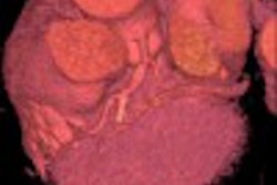
The subjects, who were considered to have an elevated risk of colorectal cancer, underwent virtual colonoscopy followed by conventional colonoscopy as the reference standard. Sensitivity was assessed on a per-patient basis, while specificity was measured both per polyp and per patient. Prone and supine CT data were acquired using collimation of 4.25 mm, pitch 1.25, 25-70 mAs,and 120 kVp.
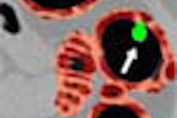
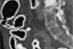
Among the patients in the study, 78 actually had polyps -- a total of 309 lesions –- while 60 were clear of the growths. The radiographers pinpointed 140 lesions -- about 45%; the radiologists noted 145 of the lesions -- about 47%, said Thomassen-de Graaf.
The only major difference was in identification of large lesions greater than 10 millimeters, she said. The radiographers identified 10 of these lesions, or 77% of the total, compared with the radiologists who discovered 12 of the large growths or 92% of the total. Still, the work radiologists excelled at is essential, inasmuch as lesions 10 mm and larger are considered clinically significant.
Neither the radiologists nor the radiographers did well in determining if the patients had no disease. The overall sensitivity was 15% for the radiographers and 19% for the radiologists. Their sensitivity increased for larger lesions – reaching 61% for the radiographers and 67% for the radiologists if the precancerous growths were 5-9 mm in size and 91% and 94% respectively if the lesions were 10 mm or larger.
“Thank God the radiologists did a little bit better,” said Dr. Bernard Birnbaum of New York University Medical Center, a co-chairman of the sessions where Thomassen-de Graaf presented her findings. Birnbaum suggested that in the litigation-conscious United States the use of radiographers in making diagnoses might be limited.
“Perhaps we need a new breed of person who would fall between a radiographer and a radiologist to perform this type of work,” suggested co-chair Dr. Janet Husband of Royal Marsden Hospital, London, United Kingdom. Thomassen-de Graaf said work is underway in Amsterdam to train radiographers as paramedical diagnosticians.
By Edward Susman
AuntMinnie.com contributing writer
December 2, 2002
Copyright © 2002 AuntMinnie.com