PHILADELPHIA - The Society of Nuclear Medicine selected a series of images from a study conducted by researchers at the Hamamatsu Medical Imaging Center in Hamakita, Japan, the University of Washington in Seattle, and the University of Michigan in Ann Arbor, MI, as the winner of Image of the Year at its annual meeting. The award was announced Monday by Dr. Henry Wagner, director of Radiation Health Sciences at Johns Hopkins in Baltimore.
The study, "Brain FDG PET Imaging in a Population-Based Cohort of Asymptomatic Subjects: Initial Findings," was led by Dr. Satoshi Minoshima, professor of radiology and bioengineering, head, primate PET imaging suite, at the University of Washington.
"I selected the image because it best represents the theme of this year's meeting, which is creating lifetime images of health and disease," Wagner said.
Wagner introduced the selection of the images by sharing his vision of a healthcare system of the future. In this system, everyone will have a periodically updated chip containing lifetime manifestations of his or her state of health, Wagner said.
"The patient's personal physician will enter the patient's chip into the database of disease manifestations," he said. "Abnormal manifestations in the person's database will be identified, and possible therapeutic courses of action suggested."
According to Wagner, the health chip will search an international health manifestation database (IHMD) every three months to answer the following questions:
- Is anything wrong?
- What is going to happen?
- What can be done about it?
- Is the treatment helping?
- How did it happen?
"Rather than trying to give a name to a patient's disease, putting him or her in a disease 'box', the person's health chip will reveal all the manifestations of the patient's health and illness," said Wagner.
The health manifestations on the patient's chip will search the IHMD to characterize the illness, predict what is likely to happen, and suggest possible treatments, he said. To create an IHMD, Wagner said that well-defined subsets of images of disease manifestations should be extracted from literature and personal Web sites.
By creating such an entity, physicians will be able to compare the patient's anatomical, functional, and biochemical imaging studies to images in the IHMD by precise overlaying or co-registrration of the images, Wagner maintained.
 |
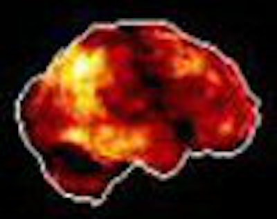
| Disease detection using 3D-SSP PET and MRI, normal database: Z-score maps from the Alzheimer's disease vs. normal database. If the patient's FDG images are most similar to these database images, the patient may have Alzheimer's disease. Image courtesy of the Society of Nuclear Medicine and Dr. Satoshi Minoshima. |
According to the researchers, early detection of brain disorders facilitates patent enrollment in therapeutic trials. Their study was designed to evaluate brain glucose metabolism, longitudinally, in a population-based cohort of asymptomatic subjects.
For the image-award winning study, the scientists acquired whole-body and whole-brain PET images from 551 asymptomatic subjects as part of the on-going study. The patients included 147 women (mean age, 45) and 404 men (mean age, 42).
PET images were acquired for 20 minutes starting one hour after FDG injection using a scanner that collected 230 slices (3.2 mm slice thickness) over a Z-axial distance of 732.8 mm.
The researchers wrote that brain gray matter activity was extracted using 3D stereotactic surface projections (3D-SSP) and normalized to global and pontine activities. Age-associated changes and gender differences were investigated using pixel-wise regression and T statistics. Metabolic abnormalities in individual cases were assessed using Z-score maps generated by age-adjusted regression, they reported.
"The most significant age-associated metabolic decline (Z=12.3, 0.4% decline/year) was seen in the medial frontal cortex, bilaterally extending to anterior cingulate and lateral frontal association cortices as well as the operculum and superior parietal and anterior temporal cortices," wrote the authors.
They also found, when adjusted for age, significantly greater metabolic activity in the frontal and anterior cingulate cortices in women and cerebellar hemisphere and vermis in men. The researchers wrote that focal metabolic reductions were seen in 11%, 12%, and 17% as well as diffuse frontal hypometabolism of 6%, 9%, and 3%, of subjects’ less than 40 years, 40-55 years, and greater than 55 years, respectively.
Cerebellar hypometabolism did not seem to be age-specific, as it was seen in 5% of each group. However, they said that Alzheimer's disease patterns were seen in 0.4% of patients less than 55-years old, in contrast with 7% of those patients older than 55.
The group concluded that their ongoing study has shown that analysis of brain glucose metabolism in a population-based cohort showed age and gender associated differences. In addition, focal or frontal hypometabolism was seen frequently in asymptomatic subjects and Alzheimer changes increase with age.
"The pathophysiologic significance of these findings will be determined in a longitudinal follow up," the authors wrote.
By Jonathan S. BatchelorAuntMinnie.com staff writer
June 22, 2004
Related Reading
LSO PET/CT study wins Image of the Year, June 24, 2003
PET, SPECT studies chosen as SNM images of the year, June 18, 2002
Wagner sees bright future for molecular imaging 10/31/02
Wagner taps PET Alzheimer's study as SNM 2001's Image of the Year, June 26, 2001
Copyright © 2004 AuntMinnie.com