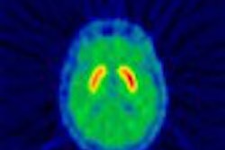
Using a "truly simultaneous" combination of functional transcranial Doppler (fTCD) sonography and H215O PET imaging, German researchers have identified differences in cerebral function that address the working-memory impairment experienced by schizophrenics.
The researchers sought to link the spatial information provided by PET with high-resolution temporal data from fTCD to pinpoint cerebral blood flow. Theirs is the first reported use of PET-fTCD to study cognitive activation in schizophrenics, according to a study published in the Journal of Nuclear Medicine.
Working-memory problems are a devastating aspect of schizophrenia, directly affecting the patient’s ability to learn skills and function in the community.
During the pretesting of their simultaneous imaging combination, the researchers noticed different activity levels around the anterior cerebral arteries (ACA) in schizophrenic versus control subjects. In addition, earlier PET studies suggesting a connection between cinguloparietal dysfunction and working-memory impairment and fTCD of the middle cerebral arteries (MCA) had shown promise for studies of the brain at work.
Although continuous insonation of the MCA over several minutes is much easier technically, the German group decided to focus on a comparison of PET and fTCD findings in the ACA.
"To our knowledge, there is no report in the literature of fTCD of the ACA in schizophrenia and of the role of fTCD of the ACA in working memory in healthy subjects," wrote Dr. Osama Sabri and colleagues from Aachen University of Technology, the University of Leipzig, Technical University Munich, and Klinikum Chemnitz in Germany. "Therefore, a truly simultaneous PET-fTCD acquisition can close this gap because PET can serve to cross-validate the findings of fTCD in the ACA" (JNM, May 2003, Vol. 44:5, pp. 671-681).
Cerebral blood-flow velocity patterns of both anterior cerebral arteries were observed using a 2-MHz pulsed-Doppler device (Pioneer TC4040, Nicolet EME, Kleinostheim, Germany) and 3-D PET imaging (ECAT Exact 922/47 scanner, Siemens Medical Solutions, Erlangen, Germany) after a bolus injection of H215O, or radioactive water.
The researchers examined 11 clinically stable chronic schizophrenics who were able to perform working-memory tasks, and 10 control subjects who were matched for age, sex, education, and IQ. To engage their working memory, the subjects were asked to observe a random sequence of single-digit numbers and press a button whenever the number presented was the same as that presented two slots back in the sequence.
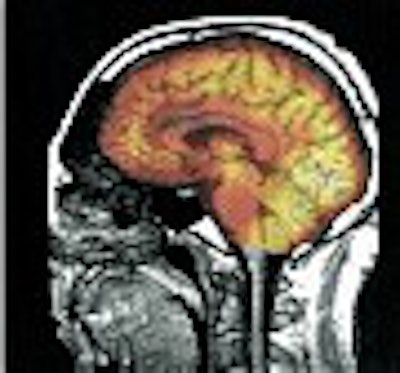
Below, an illustration of activation clusters. Top left, the sagittal averaged PET image of four scans under activation condition. Bottom left is the overlay of an averaged PET image onto the individual MRI dataset. The top right image depicts the identification and delineation of significant activation clusters. The clusters were projected into counter of PET image for better visualization. The same procedure was done for significant deactivation clusters in the bottom right image.
 |
| Reprinted by permission of the Society of Nuclear Medicine from: Sabri, O et al. A Truly Simultaneous Combination of Functional Transcranial Doppler Sonography and H215O PET Adds Fundamental New Information on Differences in Cognitive Activation Between Schizophrenics and Healthy Control Subjects. Journal of Nuclear Medicine 2003, Vol. 44, pp. 671-681. |
This "two-back" task requires a continuous updating in the working memory, yet there was no significant difference in error rates or reaction times between the two groups of subjects. The authors credited their criteria for selecting clinically stable schizophrenics for avoiding the "strongly pronounced positive symptoms" that correlate with impaired performance on memory tests.
Nevertheless, differences were seen between the schizophrenic and control subjects in the data collected by PET-fTCD.
As had been noted in the pretesting phase, the schizophrenic patients showed more extended significant activations in the supply area of the ACA than did the control subjects while using their working memory. Meanwhile, blood-flow changes could be correlated with shorter reaction times for the controls but not the schizophrenic subjects.
"The fact that our schizophrenic patients showed significantly more activations in the supply area of the ACA and left temporal areas (with a task performance comparable to that of control subjects) could be interpreted as recruitment to compensate for partial neural dysfunction," the authors wrote. "However, the fact that only healthy control subjects show a good correlation between task performance and blood flow-dependent imaging parameters indicates a qualitative difference in cognitive processing between control subjects and schizophrenic patients."
While healthy subjects were capable of adapting and increasing their cerebral blood flow to meet their needs, the schizophrenic patients instead activated a much greater number of neurons from the start.
"All facts could be interpreted as a sign of alternative, less-efficient problem-solving strategies in schizophrenia, less efficient than those used by healthy control subjects, which lead to the working-memory deficits observed during the further course of this disease," Sabri and colleagues wrote.
By Tracie L. ThompsonAuntMinnie.com contributing writer
June 2, 2003
Related Reading
MRI separates schizophrenia, mood disorders in adolescents, December 16, 2002
MRI findings predict development of symptomatic psychosis, December 11, 2002
In vitro MRI findings support vascular cause of late-life depression, October 18, 2002
Prefrontal cortical activity and dopaminergic function linked in schizophrenia, February 4, 2002
Copyright © 2003 AuntMinnie.com