LOS ANGELES - The Society of Nuclear Medicine chose images from a German PET screening study and a U.S. SPECT study of animal models as winners of Image of the Year at its 49th annual meeting.
The awards were announced by the past chairman of the society’s scientific program committee, Dr. Peter Conti. Conti, who directs the University of Southern California's PET Imaging Science Center in Los Angeles, said it was the first time two images have been selected to share the honor.
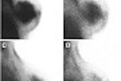
The PET study was conducted by researchers at the Hospital of Johann Wolfgang-Goethe University in Frankfurt, Germany. The investigators sought to evaluate the use of F-18-FDG-PET for screening malignancies in patients with paraneoplastic syndromes (PS). These syndromes comprise a variety of clinical symptoms and diseases associated with underlying malignancy. The group used the modality to assess patient management by differentiating the extent of the disease.
 |
| A patient diagnosed with dermatomyositis (shown by the red arrows) also exhibits bronchogenic carcinoma (shown by the blue arrow) on a screening PET scan. Image courtesy of the Society of Nuclear Medicine and the Hospital of Johann Wolfgang-Goethe University. |
Over the course of five years, the team evaluated 30 patients (17 males and 13 females averaging 55 years old) with suspected PS. Diagnoses on the patients ranged from cerebellar degeneration, erythrodermia, dermatomyositis, polneuropathia, and others.
PET scans were conducted and reviewed for convincing signs of malignant metabolism, and the results were compared to histopathologic, radiologic, and follow-up data. The group detected underlying malignancy in 8 patients, of which PET found 7 of the 8 malignant neoplasms. On the basis of cost and time needed to diagnose the malignancies with conventional tools, the group said it found F-18-FDG-PET to be of reasonable value as a screening tool in suspected PS.
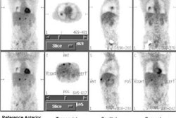
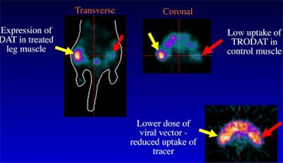
A team from the University of Pennsylvania in Philadelphia shared the top honors this year with its research on small-animal functional imaging using ultra-high-resolution SPECT. The group used the modality to perform quantitative studies of dopamine transporters.
 |
| A series of images of 99mTc Trodat-1 in a mouse leg muscle measured using ultra-high-resolution SPECT shared the Image of Year award. Image courtesy of the Society of Nuclear Medicine and the University of Pennsylvania. |
Four male mice were injected with 99mTc Trodat-1. They were scanned on a SPECT system at a spatial resolution of 0.83 mm at a 3-cm radius of rotation using pinhole collimators. This technique enabled the team to achieve an image resolution of less than 1 mm, according to Conti.
Each mouse underwent two studies, and a reference tissue kinetic modeling analysis of the time-activity data in the striatum and cerebellum was used to quantify the availability of dopamine transporters. In addition, the group conducted an equilibrium ratio of striatum to cerebellum to obtain another measure of dopamine transport binding.
The SPECT data was then compared with ex vivo biodistribution data from the striatum and cerebellum. The researchers reported excellent correlation between the ex vivo data and the kinetic studies. Because the results were so accurate, the group sees the possibility of further studies of animal models of human disease, particularly cerebral binding sites in mice, using its pinhole SPECT technique.
By Jonathan S. BatchelorAuntMinnie.com staff writer
June 18, 2002
Related Reading
Wagner taps PET Alzheimer’s study as SNM 2001’s Image of the Year, June 26, 2001
Copyright © 2002 AuntMinnie.com