Armed with a shiny new 16-slice ECG-gated scanner and a heart phantom, German researchers set out to optimize their coronary artery calcium (CAC) evaluation protocol. They found that using 3-mm slice widths and submillimeter collimation enables the detection of smaller calcium deposits than with 3-mm slices and 1.5-mm collimation. The results also showed that calcium mass is the most stable measurement of plaque burden across multiple protocols.
At the 2002 RSNA meeting in Chicago, Bernt Ohnesorge, Ph.D. and colleagues from Siemens Medical Solutions in Forchheim, Germany, discussed their evaluation of two examination protocols for coronary artery calcium scoring.
The latest generation of 16-slice CT scanners enables acquisition of the entire heart (120 mm) in a single breathhold with relatively short exposure times. Submillimeter collimation is also possible using longer exposure times and a slightly higher radiation dose, Ohnesorge said, but has not been shown to offer significant additional benefit. The study was undertaken in an effort to assess the utility of submillimeter collimation.
"We investigated the scan protocols for a new 16-slice scanner in a phantom study by means of the optimized sensitivities for small lesions, as well as the consistency of measurements," Ohnesorge said. The experiments were conducted on a Siemens Somatom Sensation 16.
The group investigated two protocols, with one crucial difference between the two: one used a 16 x 1.5-mm collimation and the other, 16 x 0.75-mm collimation. Both protocols relied on ECG-pulsed spiral acquisition, ECG-controlled dose modulation, 0.42-second rotation speed, pitch 3.7, 120 kVp, and 100 mAs. The table speed was 13.2 mm/sec for the 1.5-mm collimation and 6.6 mm/sec for the 0.75-mm setting.
The images were reconstructed in diastole using 40% overlap with 1-mm and 2-mm slice widths for 1.5-mm collimation; and with 2-mm and 3-mm slice widths for 1.5-mm collimation, according to Ohnesorge. Temporal resolution was 105-210 ms.
The results were quantified in terms of calcium volume equivalent and mass using calcium thresholds (low-, medium- and high-density) and individual calibration factors, as well as Agatston score normalized to slice overlap.
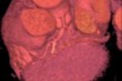
The group used a commercially available phantom with precisely measured calcium-density levels to test the protocols. Data were also acquired in 20 patients -- 10 for each protocol -- but these results were included only anecdotally.
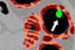
With the calcium phantom, the group found that using 0.75-mm collimation versus 1.5-mm collimation yielded longer scan times (40 sec vs. 20 sec) and a slightly higher radiation dose (1.7 mSv vs. 1.5 mSv). The smallest detectable calcifications couldn't be seen with the 3-mm collimation, Ohnesorge said.
"When we look at the sensitivity to find small lesions, we see a clear difference," he said. "(As for) the very smallest lesions, we actually only identified those of lower density based on the thin-slice collimation protocol. The sensitivity of small lesions is definitely higher using thinner collimation. As we know from the helical CT world, (using) the same slice width with thinner collimation improves the slice sensitivity profile under temporal resolution," he said.
But while longitudinal resolution was better using 1-mm and 2-mm slice-width reconstructions, the detection of calcifications could not be improved due to signal-to-noise ratio, he said.
"In the higher-density plaques, we find that with different collimations all the way down to 1-mm slices, that (calcium) volume and Agatston scores tend to be overestimated and increase with larger slice width," Ohnesorge said. "(With) medium-density plaques, volume tends to increase with larger slice widths, while total mass and Agatston scores seem to be not affected."
In low-density plaques, calcium volume and total mass are largely unaffected by different collimations, but Agatston scores tended to increase, possibly due to partial-volume effect that suppresses part of the lesion, Ohnesorge said.
"We see that the new 16-slice scanners can provide single-breathhold acquisition with different slice collimations," he said. Agatston score and calcium volume appear to be strongly dependent on slice width and slice collimation, while calcium mass is largely independent.
"The scan protocols for thin-based collimation can provide higher sensitivity to detect smaller calcified lesions, so in conclusion, quantification of calcium mass is most consistent with different scan protocols, and should become the quantification method of choice in the future," Ohnesorge said.
In response to a question from the audience, Ohnesorge said the patient data is consistent with results using the phantom, and will be quantified in an upcoming study. For now, his group recommends using 3-mm slice widths and thin collimation for optimal results. Using thinner slices could potentially uncover even more calcium, he said, but the benefits of doing so have not been evaluated.
By Eric BarnesAuntMinnie.com staff writer
December 18, 2002
Related Reading
Population-based study links coronary calcium and mortality, December 1, 2002
Technology keeps CT vital for coronary assessment, November 13, 2002
Coronary calcium, C-reactive protein predict cardiovascular event, October 3, 2002
Copyright © 2002 AuntMinnie.com