VIENNA - A study presented at today's European Congress of Radiology meeting found distinct advantages in performing digital mammography on a flat-panel digital mammography system versus a computed radiography-based digital mammography unit. While researchers gave CR mammography good marks for image blackness, the flat-panel system outperformed in all other categories.
Recent studies have demonstrated that flat panel-based FFDM is at least the equal of conventional film-screen systems, and in some cases is superior, according to Dr. Georg Pfarl of the University of Vienna. Digital mammography systems based on digital storage phosphor CR have not been studied in great detail, however, and the Vienna researchers embarked on a study to compare a CR digital mammography system to a flat-panel unit.
The prospective study involved 152 patients with a mean patient age of 57 years, examined between October 2001 and March 2002. The group used a Senographe 2000D FFDM system (GE Medical Systems, Waukesha, WI) and printed film to a wet laser printer (Agfa HealthCare, Mortsel, Belgium).
The CR unit was a Mammomat 3000 Nova (Siemens Medical Solutions, Erlangen, Germany) outfitted with FCR 5000MA storage-phosphor plates (Fujifilm Medical Systems, Tokyo, Japan). The CR mammography images were printed to a Fuji dry laser printer.
Images were reviewed by a panel of five radiologists, with image quality ranked on a scale of 1-5, with 1 being "excellent." Image quality criteria was rated in 11 categories, including contrast, sharpness, blackness, artifacts, skin line visibility, image noise, and detection and magnification of microcalcifications.
Flat-panel digital mammography was given higher marks in 10 of the 11 categories, with CR mammography getting higher ratings only in the image blackness category, Pfarl said. The overall image quality rating was 1.49 for flat-panel and 2.01 for CR mammography.
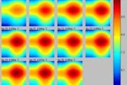
Pfarl showed a series of comparisons between flat-panel and CR mammography images. Flat-panel images demonstrated more visible skin line than CR mammography, and microcalcifications were better visualized, he said. Another image demonstrated better visualization of the borders of an uncircumscribed mass.
Pfarl concluded his talk by stating that image quality on a flat-panel unit with cesium iodide/amorphous silicon detectors was superior to that of CR-based digital mammography.
In the question-and-answer session, one presentation attendee pointed out what he claimed was a flaw in the study, in that it used a wet laser printer to produce flat-panel images and a dry laser for the CR mammography films. The attendee said that in his experience dry lasers are inferior to wet laser printers, and that the group should soften its conclusion due to this difference.
Pfarl acknowledged the potential for bias, and said that the group wanted to review images directly on each system's workstation, but the Siemens workstation was not ready for clinical use at the time of the study.
The study's senior author, however, Dr. Christopher Riedl, said that his group presented research last year that indicated no difference between the image quality of wet and dry lasers for digital mammography.
In another presentation during the same session, Riedl provided ECR attendees with an update on a study, first presented at the 2002 RSNA meeting in Chicago, on the use of FFDM to lower radiation dose during quality control mammograms to verify biopsy marker placement.
The University of Vienna group made 100 placements of either hookwire or clip markers in 76 patients with 88 lesions. One group received quality control mammograms at 50% of normal dose as measured in mAs; the second group at 75% of normal dose.
The group then examined how the lower settings affected average glandular dose (AGD) and image quality, with the latter measured by the visibility of both marker and lesion. The first group received an AGD reduction of 60%, while the second group had an AGD reduction of 79%. In all cases, the markers were visible, while lesions weren't visible in only six cases (one in the 50% dose reduction group and five in the 75% group).
Another group, from the University Medical Center Nijmegen in the Netherlands, compared the image quality of FFDM exams printed on hard-copy film to that of conventional film-screen mammograms. Ten radiologists reviewed images from 32 patients who had abnormalities first detected either with an FFDM unit (Senographe 2000D) or a film-screen system. Images were output to a DryView 8600 laser printer (Eastman Kodak, Rochester, NY).
The images were rated on a 10-point malignancy scale, and results analyzed with receiver operating characteristic (ROC) curve methodology. Readers viewing FFDM images scored an average of 0.85, while those viewing film-screen images scored 0.80. The group found the improvement to come from better characterization of benign masses, according to the study's presenter, Dr. Nico Karssemeijer.
Related Reading
CAD mass markers aid in reduction of mammographic interpretation errors, March 7, 2003
No compromise in breast image quality with lower FFDM dose, December 13, 2002
Copyright © 2003 AuntMinnie.com