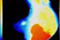
VIENNA - In a comparison of gadolinium contrast-enhanced MRI and 123I-IMT SPECT for the preoperative grading of malignant gliomas, MRI fared slightly better, but both modalities proved efficient for noninvasive imaging.
Dr. Karesten Papke from the department of clinical radiology at the University of Munster in Germany presented the results of his group’s study at the European Congress of Radiology on Sunday.
"123I-IMT is a radiolabeled amino acid that is transported into cells by an amino acid carrier system. Its uptake in the glioma correlates with proliferative activity," Papke said.
For this study population, 48 patients with untreated gliomas were studied. Gd-enhanced MRI exams were performed on a Magnetom Vision 1.5-tesla unit (Siemens Medical Solutions, Erlangen, Germany) with T1-weighted sequences before and after the administration of gadolinium. Tumor grading was based on nine criteria, including signal heterogeneity, presence of necrosis, hemorrhage, border definition, flow void, and degree of contrast enhancement.
123I-IMT (123Iodine-a-methyl tryosine) SPECT studies were conducted on a triple-head Multispect 3 gamma camera (Siemens) equipped with medium-energy collimators. Uptake of the radionuclide was quantified as the ratio between amino acid uptake in the tumor over that of the uptake in the contralateral hemisphere.
The final tumor grading was proven histologically. Out of the 48 patients, 15 tumors were characterized as grade II (based on the World Health Organization’s tumor classification system), nine tumors were grade III, and 24 tumors were grade IV.
In differentiating high-grade gliomas from low-grade ones, Gd-enhanced MRI had a sensitivity of 80%, a specificity of 91%, and an accuracy of 88%. In comparison, SPECT had a sensitivity of 67%, a specificity of 88%, and an accuracy of 81%.
"You can see that the sensitivity of MRI is slightly higher than SPECT," Papke said. "However, we still use SPECT in our practice because it provides information that gadolinium-enhanced MRI cannot, such as additional information on the size of the tumor." SPECT scans also serve as a reference should the patient require later imaging for possible recurrence, he added.
Attendees at this neurology and tumor session asked Papke if his group had used MRI perfusion studies or MR spectroscopy for grading gliomas rather than SPECT. Papke replied that neither of those MR modalities was as effective in depicting small anaplastic areas as nuclear medicine scans and biopsy.
By Shalmali Pal
AuntMinnie.com staff writer
March 5, 2001
Related Reading
MR perfusion helps diagnose brain lesions, February 16, 2001
Diffusion MRI allows early assessment of brain tumor treatment, December 12, 2000
Click here to post your comments about this story. Please include the headline of the article in your message.
Copyright © 2001 AuntMinnie.com