Combining ultrasound imaging and clinical features can help radiologists identify malignant soft-tissue tumors for radiologists, suggest findings published September 11 in Ultrasound in Medicine & Biology.
Researchers led by Yusen Zhang from Peking University Shenzhen Hospital in China found that a model combining these features achieved high diagnostic performance in evaluating these tumors compared with using imaging features alone.
"The diagnostic model also had superior accuracy compared with the performance of radiologists, suggesting its potential in aiding radiologists in clinical decision-making in patients with soft-tissue tumors," Zhang and colleagues wrote.
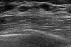
Ultrasound is the go-to imaging modality for assessing soft-tissue tumors due to its convenience and safety. With recent advancements such as color Doppler and high-frequency imaging, ultrasound also shows high resolution of superficial soft tissues.
However, the researchers noted that even with these advancements, it's still a challenge for radiologists to diagnose soft-tissue tumors with imaging or clinical information alone. They also pointed out interobserver variability and operator dependence of ultrasound as hindrances to the technology's diagnostic performance.
Zhang et al sought to test the performance of a diagnostic nomogram that incorporates both ultrasound and clinical features to differentiate between malignant and benign soft-tissue tumors. The team developed two models for its study: One blended clinical and ultrasonic features, and the other used ultrasonic features only. Clinical variables used for the combined model included patient age, tumor history, tumor location, size, boundaries, internal echogenicity, posterior acoustic changes, and blood flow patterns.
The nomograms were tested on soft-tissue tumors from various areas of the body, including the head and neck, and upper and lower extremities. Also, tumors came from the skin, subcutaneous, and muscular layers, as well as in the bone.
For the study, the authors included 613 patients with 195 malignant and 418 benign soft-tissue tumors. They also compared the performance of the models to each other, as well as that of two radiologists. They found that the nomogram that used both ultrasonic and clinical features had the highest diagnostic performance of the three methods.
| Comparison between nomograms, radiologists | ||||
| Measure | Radiologists (average) | Ultrasound-only model | Ultrasound-clinical features model | p-value |
| Area under the curve (AUC) | 0.81 | 0.89 | 0.95 | < 0.001 (for both) |
The researchers also evaluated the integrated discrimination improvement (IDI) for both nomograms. This measure reflects how the new model increases risk in events and decreases risk in non-events. The team found that the IDI between the two nomograms was 0.15 (p < 0.001), which it wrote suggested that accuracy was improved by adding clinical features.
Zhang et al wrote that they expect that the combined model may provide a reference for radiologists in classifying soft-tissue tumors that are hard to diagnose. They added that the nomogram could help reduce missed diagnoses of malignancies, as well as unnecessary biopsies.
They also called for future studies to use prospective multicenter database collection to validate the model's performance and include novel ultrasound techniques and patient follow-up to predict prognosis.
"Given the consistency of the model in variable inclusion with previous studies, we believe that the model has relatively high stability but might still be influenced by the constitution of the pathological types of soft-tissue tumors in the study population," they concluded.
The full study can be found here.