Most of the ultrasound development over the past year has been software-based, driven by the continuing growth in applications such as cranial infections, skin metastases, and sonography of all kinds with contrast agents. In addition, the portable handheld arena promises to grow tremendously across the board, in office practice as well as ER and trauma applications.

In circumstances in which conventional radiography is unavailable, such as in situations of multiple traumas or mass casualties, sonography can be used to identify internal bleeding in the chest and the abdomen, and is effective in detecting bullets or shrapnel. Portable and handheld units have found their way into triage on the battlefields of Iraq.
Ultrasound also shows increasing value for sonographically guided biopsies and therapeutic injections. Radiologists are using the modality at a higher rate, while nonradiologists, particularly in echocardiography, have also increased utilization.
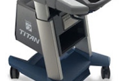
The use of 3-D ultrasound is becoming popular in both research and clinical applications, providing information that cannot be acquired with two-dimensional studies. And the equipment is finally becoming more ergonomically functional, with improved transducer shapes and control panel layout, enabling sonographers to capture a better-quality image and improve patient throughput without repetitive-stress discomfort. One vendor is even introducing a handheld remote control interface.
By Robert BruceAuntMinnie.com contributing writer
November 20, 2003