Whole-heart 4D flow and short-axis cine MRI reveal characteristics associated with impaired blood flow in the left atrial chamber of the heart, according to a study published March 7 in Radiology: Cardiothoracic Imaging.
The study findings could help clinicians better assess heart blood flow abnormalities, wrote a team led by Maurice Pradella, MD, of Northwestern University Feinberg School of Medicine in Chicago.
"We hypothesized that potential associations between [4D flow parameters and conventional volumetric-based left atrial assessment] could help to better characterize participants with impaired flow characteristics and enhance the understanding of mechanistic relationships between left atrial flow and volumetric parameters," the group noted.
Previous research has suggested that 4D flow-based assessment of blood stasis and peak velocity in the left atrium and left atrium appendage could help characterize impaired blood flow in the heart and therefore, risk of blood clot formation, Pradella and colleagues explained. But links between conventional volumetric and functional left atrium parameters with flow characteristics in the left atrium and left atrium appendage have not been explored in detail, they wrote.
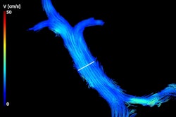
To address this knowledge gap, the investigators conducted a study that included data taken from 158 patients enrolled in Northwestern's Multi-Ethnic Study of Atherosclerosis, all of whom underwent prospective cardiac MRI between 2019 and 2020; the exams consisted of whole-heart 4D flow and short-axis cine imaging. The 4D flow MRI analysis included manual 3D segmentation of the left atrium and its appendage, and the team used these measures to quantify peak velocity and blood stasis.
The team used short-axis cine data to define left atrial contours, extracting 3D–based left atrial volume measures to calculate left atrial emptying fractions [total LA emptying fractions (LAEFtotal), active LA emptying fraction (LAEFactive), and passive LA emptying fraction (LAEFpassive)]. The group also calculated left atrial stasis, left atrial peak velocity, left atrium appendage stasis, and left atrium appendage peak velocity flow parameters to identify any associations with left atrial volume and left atrial emptying fraction.
The group found the following:
- A one-unit increase in LAEFtotal was linked to decreased left atrial stasis (p < 0.001).
- Increased LAEFactive was linked to increased left atrial peak velocity (p < 0.001).
- Increased minimum left atrial volume was most associated with impaired left atrial appendage flow and lower left atrial appendage peak velocity.
"Increased absolute and indexed minimum left atrial volume was most closely associated with impaired left atrial flow, which is a known risk factor for left atrial thrombus formation and potentially ischemic stroke," the authors concluded. "Thus, 3D-based left atrial volume quantification may be a useful surrogate measure for left atrial and left atrial appendage flow abnormalities in future studies."
The complete study can be found here.