Researchers have found that MRI reveals functional brain alterations that are associated with major depression, according to a study published February 19 in JAMA Network Open.
The study results could help clinicians better care for depressed patients, wrote a team led by Xinyi Wang of Southeast University, Nanjing, China.
"[Major] depressive disorder (MDD) is associated with reduced quality of life and increased risk of suicide and self-wounding," Wang and colleagues noted. "Understanding the neurobiological mechanisms underlying depression is a crucial aspect of developing improved therapeutic options and minimizing adverse outcomes."
Previous research using functional and structural MR imaging has explored the neurobiological mechanisms of depression and has noted depression-related alterations, such as hippocampal volume reductions and increased amygdala activity during emotional tasks. But it's still unclear "how far these alterations persist and whether prior exposure to depression affects brain function and structure later in life," the group wrote.
The study included 20,484 individuals with lifetime depression (61.7% of whom were women) and 25,462 healthy controls (55.3% of whom were men). The team took this data from a UK Biobank cohort enrolled between January 2014 and December 2018; all study participants underwent brain MR imaging. In the group with depression, the condition was evaluated by the following criteria:
- Seeking help for the condition
- Self-reported depression
- Antidepressant use
- Depression definition by Smith et al
- Hospital International Statistical Classification of Diseases and Related Health Problems, Tenth Revision (ICD-10) diagnosis codes F32 (bipolar disorder) and F33 (manic episode)
- Composite International Diagnostic Interview Short Form score
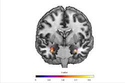
The investigators assessed the number of depression criteria study participants met, ranging from one to six. They calculated functional MRI measures using "voxel-wise fractional amplitude of low-frequency fluctuation (fALFF), global correlation (GCOR), and local correlation (LCOR)," and structural brain measures using gray matter volume (GMV).
The team found that, across all depression criteria, study participants with lifetime depression showed consistent decreases in fALFF, LCOR, and GCOR but not in gray matter volume. Those who had been identified with Hospital ICD-10 diagnosis codes F32 and F33 and antidepressant use had the most alterations in these measures.
"We found significant alterations in all functional and structural measures for most lifetime depression groups, except in patients who met all 6 criteria or met only 1 criterion when using GMV and GCOR," the group reported.
"Results of this cross-sectional study indicate that lifetime exposure to depression was associated with robust functional changes, with a more restrictive depression definition revealing more pronounced alterations," the authors concluded.
The complete study can be found here.