MRI shows variation in the size of metastasized liver lesions from colorectal cancer depending on whether hepatobiliary phase (HBP) or dual contrast MRI exams are used, researchers have found.
The results underscore the importance of clinicians choosing one method of tracking patients with metastasized colorectal cancer and using it consistently, wrote a team led by Ruben Ngnitewe Massa'a, MD, of the University of Wisconsin at Madison. The study findings were published August 7 in the Journal of Computer Assisted Tomography.
"[The] measurement size of metastatic colorectal cancer lesions in the liver is significantly different between HBP images and dual contrast images in dual contrast liver MRI," the group wrote. "Metastatic lesions should be consistently measured on the same set of images longitudinally for accurate comparison."
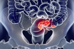
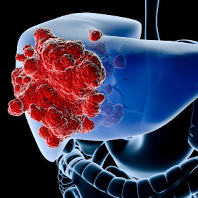
Colorectal cancer is the third most diagnosed cancer in the U.S., the investigators noted. About a quarter of colorectal cancer patients have metastatic liver disease when they are diagnosed, and the extent to which these metastases involve the liver affects patient outcomes.
Precise identification of the number and location of hepatic metastatic lesions is particularly important for clinical management," Ngnitewe Massa'a and colleagues wrote. "Prior studies have demonstrated that [MRI] with liver-specific contrast agents can detect metastases with a high degree of accuracy."
There are two different kinds of liver MRI contrast agents -- hepatobiliary (HBA) and extracellular. Typically, patients undergo a dual-contrast MRI exam protocol under which they are scanned with an HBA, then with an extracellular agent. But patients may also undergo HBP MR imaging, and some studies have shown differences in lesion measurements between the two techniques.
"This apparent variability [raises] concern for longitudinal assessment of lesions, particularly when evaluating response to therapy or determining whether surgery or embolization/ablation is indicated," the investigators noted.
To explore variations between HBP and dual-energy MRI liver lesion measurements, the team conducted a study that included 70 liver lesions in 39 patients with known colorectal carcinoma who had undergone both HBP and dual contrast liver magnetic resonance imaging. The lesions were measured on both HBP and dual contrast images with software that automatically delineated the lesion edge, and thus, the lesion diameter. The investigators used software to compare lesion measurements from the two different MRI exams. The lesions were also measured by two abdominal radiologists.
The group reported that HBP MRI measurements were larger than those found on dual contrast MRI, both by the software and by the radiologist readers. This result was statistically significant (p < 0.001).
| Liver lesion size by type of MRI exam and reader | ||
| Type of exam | Software | Radiologist readers |
| HBP MRI | 10.9 mm | 10.5 mm |
| Dual contrast MRI | 10.5 mm | 9.6 mm |
The findings highlight the importance of choosing a method for measuring colorectal cancer liver metastases and using it consistently, according to the authors.
"Both software-based and radiologist-based measurements of colorectal cancer liver metastases are significantly larger on HBP than dual contrast images," they concluded. "Based on these findings, we recommend that longitudinal assessment be performed consistently on either HBP or dual contrast phases to avoid introduction of avoidable variability."
The complete study can be found here.