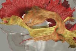
A diffusion-tensor MR image (DTI-MRI) of a mouse kidney described as "stunning" has been named the top image in the second annual "research in progress" photo competition held by BMC, the scientific publisher formerly known as BioMed Central.
The image was acquired using DTI-MRI by Nian Wang, PhD, assistant professor of radiology at the Center for In Vivo Microscopy at Duke University. The image demonstrates a mouse kidney at 10-micron resolution and has been colorized to represent the orientation of different tubules in the organ that collect filtrate from blood passing through the kidney and process it into urine.
 Diffusion-tensor MR image of a kidney at 10-micron isotropic resolution. The image makes it possible to estimate location, orientation, and anisotropy of the tubular tracts; colors represent the different orientation of tubules. Image courtesy of BMC.
The Duke center focuses on developing novel MRI methods for detecting tissue microstructures, according to a statement released by BMC. Researchers at the center have found that the nondestructive nature of MRI and its ability to assess renal microstructure in 3D make the modality a promising tool for understanding complex structures.
The image represents the ability of science and research to offer new perspectives on life and shows how unexpected beauty can be found almost as a side effect of a researcher's work, according to Rachel Burley, publishing director of BMC and SpringerOpen.
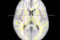
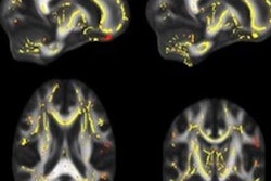
The winning image was selected from among 373 entries, according to BMC. The runner-up was a 3D reconstructed image of brain circuitry structure in a common fruit fly using synchrotron x-ray tomography.