The ability of diffusion-weighted MRI (DWI-MRI) to differentiate between ocular melanoma and retinal detachment can help direct the appropriate proton beam therapy for patients, according to a study presented at this month's European Congress of Radiology (ECR).
German researchers found that DWI-MRI detected retinal detachment in almost two-thirds of patients in the study, with apparent diffusion coefficient (ADC) values showing a significant difference between ocular melanoma and retinal detachment.
In her ECR 2013 presentation, lead study author Dr. Katharina Erb-Eigner, from the Institute of Radiology at Charité Medical University in Berlin, noted that DWI-MRI has been used primarily for neuroradiological applications to investigate conditions such as stroke and brain abscesses. More recently, DWI has branched out to oncology to characterize lesions of the head and neck, breast, liver, and prostate.
Ocular melanomas are the most common intraocular tumors in adults, and ocular MRI is utilized to assess the extent of a lesion.
"At our institution, patients with large ocular melanomas get proton beam therapy, and we use ocular MRI to plan the proton beam therapy," Erb-Eigner said.
The problem for radiologists is that it can be difficult to distinguish ocular tumors from retinal detachment with noncontrast and contrast-enhanced T1- and T2-weighted MRI sequences due to retinal effusion. Most patients with ocular tumors present with retinal detachment, according to the researchers.
"The signal of the retinal detachment and the tumor may be similar, which may be due to hemorrhage on the detachment and to the variable content of the ocular melanomas," Erb-Eigner explained. "So, in some cases, we discovered difficulties to distinguish the ocular melanoma from the retinal detachment."
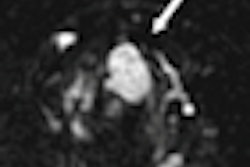
The MRI scan involves the use of a small surface coil that is positioned over a patient's eye, which is closed during the exam. A head coil is also applied for diffusion-weighted MR imaging.
 |
| A small surface coil is placed over the patient's eye, which is closed during the scan. A head coil is also used for diffusion-weighted MR imaging. All images courtesy of Dr. Katharina Erb-Eigner. |
The study included 30 consecutive patients with ocular melanoma and utilized either 1.5-tesla or 3-tesla MRI. The conventional imaging protocol included T1- and T2-weighted sequences, as well as contrast-enhanced T1-weighted sequences.
DWI was added to the conventional ocular MRI protocol, with researchers placing a region of interest on the DWI results for both ocular melanoma and retinal detachment. They then calculated ADC values.
In total, 23 (61%) of the 30 patients were found to have retinal detachment. The mean ADC value for ocular melanoma was 922 x 10-6 mm2/sec, which differed significantly from the mean ADC for retinal detachment of 2,035 x 10-6 mm2/sec.
 |
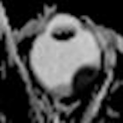
| In the noncontrast T1-weighted image (left) and T2-weighted image (center) there is an intraocular mass, but it's hard to discern if there is both a tumor and retinal detachment. With DWI (right), the tumor is much more visible and the area can be targeted for proton beam therapy. |
Based on the results, Erb-Eigner and colleagues concluded that DWI-MRI helps to delineate ocular melanoma from retinal detachment. "Since the acquisition time of DWI is very short, we recommend including DWI in ocular MRI, if ocular mass is suspected," she added.
DWI-MRI has become the imaging technique of choice for ocular melanoma and retinal detachment at Charité. Currently, about one patient per week undergoes the procedure, but Erb-Eigner expects that rate to change.
"We are starting to use it more and more," she said. "In some cases, it is very clear what is retinal detachment when there is no hemorrhage. In the cases where it is not clear, we use the DWI technique to delineate the tumor."