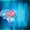
Functional MR images (fMRI) of the brain indicate that a condition known as leukoaraiosis -- once thought to be harmless -- can adversely alter brain function in elderly individuals and decrease their language and verbal skills, according to a study published online August 13 in Radiology.
In an age-matched comparison with healthy individuals, researchers at the Mayo Clinic in Rochester, MN, detected larger clusters of leukoaraiosis lesions in the brain regions associated with language and verbal function in individuals with the condition.
"The results of our experiment add to growing evidence that leukoaraiosis is not a benign manifestation of aging but rather an important pathologic condition that alters brain function," concluded lead author Dr. Kirk Welker, assistant professor of radiology at Mayo, and colleagues.
Not so benign?
Leukoaraiosis, also known as small-vessel ischemia or arteriolosclerosis, is a condition in which diseased blood vessels lead to small areas of damage in the white matter of the brain. The lesions are frequently detected in the brains of people older than 60 and are particularly prevalent in individuals with cerebrovascular disease, hypertension, or diabetes (Radiology, August 13, 2012).
Mayo's prospective study recruited cognitively healthy subjects between 2006 and 2010 from the Mayo Clinic Study of Aging, which includes approximately 2,300 randomly selected individuals with no signs of dementia. These individuals had all received fMRI brain scans during the preceding year.
Welker and colleagues examined 20 individuals with a leukoaraiosis volume of 25 cm3 or more, in addition to 20 healthy control subjects who had a leukoaraiosis volume of 5 cm3 or less. The participants with leukoaraiosis were matched by age with a healthy counterpart who was no more than three years older or younger. The process resulted in a total of 18 age-matched pairs for analysis.
The researchers used an optical projector with a head coil-mounted projection screen to stimulate the brain and record functional MR images. An eye camera also was used to monitor ocular motion as each individual performed his or her series of tasks.
Functional tasks
The mental and visual exercises included word pairs, in which the first word was a category and the second word was an example of that group. For example, researchers paired the words "fruit" and "apple," which subjects would identify as a correct combination, or "dish" and "stick," which would be an incorrect relationship. A total of 26 word pairs were correct and 14 combinations were incorrect.
Participants viewed the word pairs every six seconds during 30-second segments and were asked to respond "yes" when the pairings were correct and "no" when they were incorrect.
For control purposes, the researchers also used a visual decision task in which sets of four vertical or diagonal lines were displayed together. Subjects were asked to respond "yes" when all four lines were identical and "no" if any of the lines were angled differently.
"These control stimuli created visual perceptual decisions that approximated the visual input from the semantic stimulus word pairs without providing language input," the authors explained.
fMRI evaluation
Whole-brain functional imaging was performed on a 3-tesla MRI scanner (Signa, GE Healthcare) with an eight-channel head coil. Welker and colleagues then analyzed leukoaraiosis volume scores and ages for mean, median, range, and standard deviation. The difference in age between the two groups was not statistically significant; the average age of the control group was 79 years, compared with 79.7 years for the leukoaraiosis subjects.
The researchers found no statistically significant difference between the two groups in terms of the average percentage of correct responses to the tasks. This occurred despite the fact that average leukoaraiosis volumes were quite different, with the control group showing an average volume of 0.8 cm3, compared with 35.6 cm3 for individuals with leukoaraiosis.
Although the performance scores for the two groups were the same, the researchers detected changes between them regarding brain activation while performing the tasks. Individuals in the leukoaraiosis group had reduced brain activation on fMRI in areas associated with semantic decisions, while activation was higher in areas associated with visual perceptual decisions.
There was no activation of the left posterior cingulum or the left posteroinferior temporal lobe in the leukoaraiosis participants, and there were also much smaller clusters of activation in the left inferior frontal and middle gyral regions, as well as the anterior aspect of the left superior frontal gyrus. Leukoaraiosis participants showed increased activation in the right parietal lobe and posterior right temporal lobe during visual tasks.
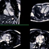
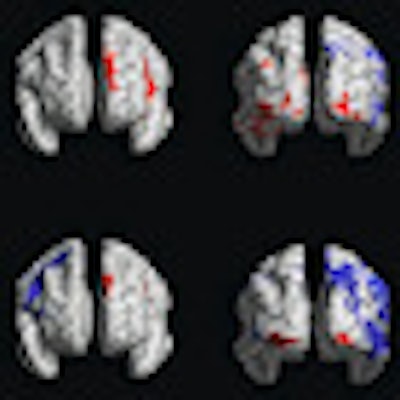
 |
| Regions of the brain in which visual perceptual decisions generated increased blood-oxygen-level-dependent (BOLD) signal compared with semantic decisions in participants with leukoaraiosis are illustrated in blue. Image courtesy of Radiology. |
Those in the leukoaraiosis group were perhaps able to achieve performance scores similar to the control group through "compensatory measures such as synaptic reorganization or disinhibition of latent neuronal pathways," the authors speculated.
"On the group level, the results of our experiment add to growing evidence that leukoaraiosis is not a benign manifestation of aging but rather an important pathologic condition that alters brain function," Welker and colleagues wrote. "Despite semantic decision performance scores similar to those of the control group, our participants with leukoaraiosis exhibited atypical cerebral activation patterns."
Identifying leukoaraiosis in the brain is important for individual patients undergoing brain mapping for surgery or other treatments, as well as for research studies, the authors added.
For the neurological health of patients, the researchers recommended that greater efforts be taken to prevent leukoaraiosis. While aging is a known risk factor for the condition, Welker and colleagues suspect that high blood pressure may also play a role.