Using 3-tesla sodium MRI, French researchers have discovered that a buildup of sodium in specific regions of the brain could be a biomarker for nerve cell degeneration in patients with multiple sclerosis (MS), according to a study published online July 17 in Radiology.
The study from the National Center for Scientific Research (CNRS) and Aix-Marseille University in Marseille found that patients with early-stage multiple sclerosis have abnormally high concentrations of sodium in the brain stem, cerebellum, and temporal pole, while patients with more advanced disease showed sodium accumulation throughout the whole brain.
Previous research has confirmed that sodium buildup in motor areas of the brain correlate directly to the degree of disability in advanced-stage MS patients.
Lead study author Wafaa Zaaraoui, PhD, a research officer at the CNRS, and colleagues conducted 3-tesla sodium 23 (Na-23) MRI exams (Magnetom Verio, Siemens Healthcare) on 14 patients who had relapsing-remitting multiple sclerosis -- the most common form of the disease -- for less than five years, and 12 individuals with relapsing-remitting MS for more than five years. The study also included 15 age- and sex-matched healthy control subjects.
The disease duration for patents with early-stage relapsing-remitting multiple sclerosis ranged from eight to 48 months, while the disease duration for patients with advanced relapsing-remitting MS was 60 to 360 months. Patients with advanced relapsing-remitting MS also were more disabled than those with early relapsing-remitting MS.
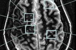
Using specially developed hardware and software, sodium MRI revealed abnormally high concentrations of sodium in specific brain regions, including the brain stem, cerebellum, and temporal pole in early-stage relapsing-remitting MS patients. Among advanced-stage relapsing-remitting MS patients, abnormally high sodium accumulation was viewed throughout the entire brain, including normal-appearing brain tissue.
 |
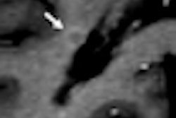
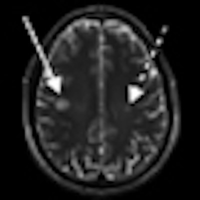
| MR images of a 33-year-old man with early relapsing-remitting multiple sclerosis shows substantial sodium accumulation in two T2 lesions with two different signal intensity patterns at T1-weighted imaging. One lesion was hypointense (solid arrows) and one was isointense (dashed arrows) to normal-appearing white matter on T1- weighted image. Images courtesy of Radiology. |
In addition, MR images showed that the amount of sodium accumulation in gray matter associated with motor skills directly correlated with the degree of disability exhibited by relapsing-remitting MS patients.
Another major result of this study is the relationship found in patients between the sodium accumulation in the gray matter compartment and disability. This finding is consistent with those of previous studies using other MR imaging modalities demonstrating the significant functional impact of gray-matter injury
Brain sodium MRI can help healthcare providers better understand the disease and monitor neuronal injury in MS patients, and possibly patients with other brain disorders, Zaaraoui and colleagues concluded. Using sodium accumulation as a biomarker of neuron degeneration also may help pharmaceutical companies develop and assess other potential treatments.