Accelerated partial-breast irradiation allows patients to receive breast-conserving radiation therapy -- traditionally a combination of lumpectomy and whole-breast irradiation with survival rates similar to mastectomy -- in five days rather than the six weeks whole-breast irradiation requires. The accelerated pace may help provide treatment for rural or elderly women, who might not otherwise have access to radiation treatment because of transportation issues.
However, the drawback of this protocol is that the treatment effect of whole-breast irradiation on occult cancer is lost. Because breast MRI has been shown to identify hidden cancer, researchers at Brigham and Women's Hospital and the Dana-Farber Cancer Institute, both in Boston, explored whether using MRI before accelerated partial-breast irradiation can determine if a patient has multifocal or multicentric disease and, therefore, help doctors plan treatment most effectively. Their findings are presented in this month's American Journal of Roentgenology (July 2008, Vol. 191:6, pp. 272-277).
Dr. Juan Godinez and colleagues studied 79 women with core needle biopsy-proven breast cancer who were eligible for breast-conserving surgery and accelerated partial-breast irradiation and who had undergone bilateral breast MRI exams. The women ranged from 29 to 75 years in age, with the mean age being 48. Twenty-nine of the 79 patients included in the study had a family history of breast cancer.
All of the MRI exams were performed with a 1.5-tesla unit (Signa HDx 1.5T, GE Healthcare, Chalfont St. Giles, U.K.) using an Invivo seven-channel, dedicated breast surface coil (MRI Devices, Waukesha, WI; Invivo, Orlando, FL).
The indications for the use of diagnostic breast MRI exam included the following:
| Indications for MRI in 79 patients with core biopsy-proven breast cancer
|
|||||||||||||||||||||||||||||
| Note -- Indications were determined by referring physician. aReferring physician's perception of young age at time of breast cancer diagnosis. bAs determined by referring physician (i.e., may include second-degree relatives). Data courtesy of the American Roentgen Ray Society. |
|||||||||||||||||||||||||||||
From 79 patients, a total of 126 MRI abnormalities were found suspicious for malignancy in the ipsilateral breasts.
For 30 of the 60 women who had core needle or surgical biopsy, the surgical plan changed to wider excision, including mastectomy; in 19 of these 30 patients, the pathology of the surgical biopsy showed further malignancy, finding 28 additional cancer foci. MRI showed 33 additional foci in these 19 patients.
 |
| Forty-eight-year-old woman with palpable lesion in right upper breast found by her physician. Mediolateral oblique (left) and craniocaudal (right) mammograms. Patient did not feel lump, and a BB marker was not placed at time of her mammogram. Images were originally interpreted as showing no evidence of suspicious lesion. However, retrospectively, possible asymmetries and architectural distortions may be present centrally (arrows). Fig 1a, b. Godinez J, Gombos EC, Chikarmane SA, Griffin GK, Birdwell RL. AJR. 2008;191(1):272-277. All images courtesy of the American Roentgen Ray Society. |
 |
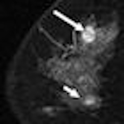
| Left, 48-year-old woman with palpable lesion in right upper breast found by her physician. Focal sonogram at site of palpable mass at 12-o'clock position shows highly suspicious irregular hypoechoic 1.4-cm mass (calipers) in posterior breast. Right, 48-year-old woman with palpable lesion in right upper breast found by her physician. Sagittal contrast-enhanced T1-weighted MR image with fat saturation shows index cancer (long arrow). Second cancer (short arrow) detected on MRI only is seen in inferior breast, 5.5 cm from incident cancer. Subsequent core biopsy confirmed that second area was another focus of grade I invasive ductal carcinoma. Fig 1c, d. Godinez J, Gombos EC, Chikarmane SA, Griffin GK, Birdwell RL. AJR. 2008;191(1):272-277. |
Godinez's team found that 49 of the 79 cases studied were appropriate for accelerated partial-breast irradiation. The results showed that MRI found more hidden cancers than physical examination or conventional imaging with mammography and sonography. Godinez and colleagues concluded that women who are eligible for accelerated partial-breast irradiation could benefit from an MRI exam before beginning treatment to ensure no hidden cancers have been missed.
By Kate Madden Yee
AuntMinnie.com staff writer
July 18, 2008
Related Reading
Solid-state scintimammography matches breast MRI, July 2, 2008
High-spatial resolution SER imaging may improve breast cancer outcomes, July 2, 2008
When MRI won't work, MDCT can help differentiate breast lesions, study suggests, June 19, 2008
Contrast MRI helps track breast cancer therapy response, June 6, 2008
Breast MRI with USPIO contrast helps assess lymph node metastases, April 25, 2008
Copyright © 2008 AuntMinnie.com