Although breast-conserving cancer therapies such as lumpectomy and radiation can be quite effective in treating the disease, invasive cancers can spread outside the primary tumor to adjacent tissue, and cancerous changes in normal-looking breast tissue have been shown to increase a woman's risk of local recurrence.
Evaluating the surrounding area of a tumor is a critical step toward a realistic assessment of a woman's prognosis and how to prevent recurrence. Dr. Jona Hattangadi and colleagues at the University of California, San Francisco assessed whether contrast-enhanced breast MRI could be a helpful tool for analyzing the microvasculature of the breast tissue beyond the tumor margin, and therefore improve local treatment options. Their work is published in the June issue of the American Journal of Roentgenology (June 2008, Vol. 190, pp. 1630-1636).
"[We based our study] on the idea that the radiologically normal-appearing nonneoplastic stroma in the breast may play a crucial role in tumor pathogenesis and response to treatment," Hattangadi wrote. "Microvessel density in tumors is known to correlate with poor prognosis in breast cancer patients, but little is known about the nature of extratumoral microvessel density and its influence on clinical outcome."
Hattangadi and her team performed signal enhancement ratio analysis of nontumor breast tissue on dynamic contrast-enhanced MR scans of 42 women. All of the patients had invasive breast cancer that had been confirmed by core biopsy or fine-needle aspiration before treatment; each woman received neoadjuvant chemotherapy for invasive breast cancer (scan one) and after one cycle of chemotherapy (scan two) between 1995 and 2002.
The median age of the study participants was 48.3, and median follow-up in the patients who remained disease-free was 52 months. Mean size of the tumor before treatment was 4.69 cm. Eighty-three percent of the study participants had invasive ductal carcinoma, 15% had lobular histology, and 1.7% had medullary carcinoma.
All of the 42 patients except one had four cycles of chemotherapy administered every three weeks, and 11 received additional weekly treatment. All patients underwent dynamic contrast-enhanced MR before chemotherapy, while 33 of the 42 patients in the study were also scanned after one chemotherapy cycle. The MR exams were performed on the ipsilateral breast only using a 1.5-tesla scanner (Signa, GE Healthcare, Chalfont St. Giles, U.K.).
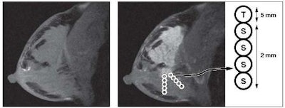
For each MR scan, signal enhancement ratio values from each region of interest (ROI) in breast tissue were characterized relative to their position, the distance from the edge of visible tumor. The mean number of ROIs for each patient was 8.5. When the team compared tumor signal enhancement ratio values with stromal signal enhancement ratio values, the mean signal enhancement ratio values from ROIs in visible tumor were higher than those in stroma.

The team found that a mean stromal signal enhancement ratio of less than 0.7 for either the scan performed before treatment or the scan performed after a cycle of chemotherapy was associated with recurrence. Univariate analysis demonstrated that pretreatment tumor size, pretreatment tumor volume, and mean stromal signal enhancement ratio at MRI scan two, or after a course of chemotherapy, were strong predictors of disease-free survival for the study participants.

"Higher mean signal enhancement ratio values in breast stroma after one cycle of chemotherapy are significantly associated with decreased local recurrence and longer disease-free survival," Hattangadi wrote. "Our findings indicate that the physiologic characteristics of the breast stroma are important factors in predicting local recurrence and clinical response to therapy."
By Kate Madden Yee
AuntMinnie.com staff writer
June 6, 2008
Related Reading
Breast MRI with USPIO contrast helps assess lymph node metastases, April 25, 2008
Reimbursement crunch throttles breast MRI CAD users, April 8, 2008
MRI beats mammography, US in detecting breast cancer in high-risk women, March 12, 2008
Catching undetected cancer with MR plus mammo in high-risk women outweighs costs, December 17, 2007
Breast MRI comparable to mammography for detecting later cancers, November 25, 2007
Copyright © 2008 AuntMinnie.com