In 2004 E. Mark Haacke, Ph.D., from Detroit's Wayne State University, discussed his work with susceptibility-weighted imaging (SWI) during a lecture at the San Francisco VA Medical Center. Haacke returns with an update on the advances of SWI in neuroimaging four years later.
Susceptibility-weighted imaging has become commonplace in today's neuroimaging protocols. SWI uses conventional high-resolution T2* gradient-echo images, phase imaging, resting state blood oxygen level-dependent (BOLD) MRI, and single voxel spectroscopy. All of these components are part and parcel of what makes SWI a powerful imaging technique.
Combining phase images with conventional T2*-weighted magnitude images offers the best of both methods. The final SWI magnitude and filtered phase images offer exquisite contrast for imaging microhemorrhages and iron content in tissue (Magnetic Resonance in Medicine, September 2004, Vol. 52:3, pp. 612-618).
SWI in the brain offers amazing contrast on 1.5-tesla imaging and even better results at higher field strength magnets (3 tesla to 7 tesla). Recent collaborations with Dr. Yulin Ge at New York University in New York City using a 7-tesla system have revealed beautiful differentiation of gray and white matter, as well as the ability to differentiate venules from tissue with SWI.
SWI's sensitivity to local changes in magnetic field means that it can detect arterial bleeds in cerebral amyloid angiopathy -- a precursor to vascular dementia in aging -- that may be as small as 50 microns. It can detect iron at the level of 10 µg Fe/gm of tissue at 1.5 tesla, or 5 µg Fe/gm of tissue at 3 telsa. SWI is very sensitive to veins because of their deoxyhemoglobin content. It's no surprise that SWI has found applications in aging, multiple sclerosis, stroke, traumatic brain injury, and tumor identification.
Aging and vascular dementia
One potential application for SWI is in Alzheimer's disease and vascular dementia. A longitudinal study done in conjunction with Loma Linda University in Loma Linda, CA, has revealed a propensity of microbleeds in a major fraction of subjects with mild cognitive impairment. When many microbleeds were present at the start of the study, these patients usually progressed to become progressively impaired or dementia cases. Several ongoing studies around the world are focusing on detecting microbleeds as a marker for aging and dementia. This work by the Loma Linda group -- under the direction of Dr. Wolff Kirsch, with analysis by the Wayne State group -- may be the first of its kind to successfully demonstrate SWI as a surrogate marker for potential vascular dementia based on microbleed detection.
 |
| SWI and aging: A connection with cerebral amyloid angiopathy. These SWI images from a 1.5-tesla scanner show a growth in the number of microbleeds over nearly two years. It is believed that these may be caused by cerebral amyloid angiopathy. This particular subject went on to develop progressive cognitive impairment (Alzheimer's disease). Image courtesy of E. Mark Haacke, Ph.D., Wayne State University, Detroit, and Dr. Wolff Kirsch, Loma Linda University, Loma Linda, CA. |
Multiple sclerosis (MS)
SWI's sensitivity to iron may open a new door into detecting lesions and the study of lesion specificity in MS.
 |
| The far left and far right images are T2-weighted and both show numerous lesions. Some are seen on SWI and some are not in the SWI filtered phase images. However, some are seen better with SWI as dark regions (high iron content), and these may eventually be shown as markers for the specificity of the disease. Image courtesy of E. Mark Haacke, Ph.D., Wayne State University, Detroit. |
Stroke and trauma
SWI offers a way to image the effects of stroke in terms of bleeding and changes in local oxygen saturation. Here, we show a case in which all other imaging methods failed to find the stroke. Very small amounts of iron can be detected with SWI. Bleeds otherwise totally invisible to conventional methods become visible with SWI.
 |
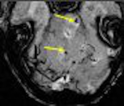
| Left, conventional high resolution (TE = 40 msec) gradient-echo magnitude image shows nothing. Right, the SWI filtered phase image shows where the stroke took place. Image courtesy of E. Mark Haacke, Ph.D., Wayne State University, Detroit. |
In trauma, SWI reveals microhemorrhages as small as a few cubic millimeters in size. Investigators at Loma Linda have detailed their successful experience with SWI in a pediatric population.
"We have used this technique in the past several years to study a wide variety of pediatric neurologic disorders," wrote Dr. Karen Tong and colleagues in the American Journal of Neuroradiology, adding that they have found it useful for detecting hemorrhagic lesions in traumatic brain injury among many other disease processes (AJNR, January 2008, Vol. 29:1, pp. 9-17).
 |
| SWI can reveal many small lesions often missed with other methods. Conventional gradient-echo images with TE = 20 msec can reveal larger hemorrhages, but are limited in the number and size of lesions seen. Image courtesy of E. Mark Haacke, Ph.D., Wayne State University, Detroit. |
NICE
To advance SWI in clinical practice as well as medical research and pharmaceutical R&D, the MRI Institute for Biomedical Research at Wayne State, along with other collaborators such as those at Loma Linda University and the Detroit Medical Center, have developed the Neurovascular Center of Excellence in Magnetic Resonance Imaging (NICE). This project was funded by a 2005 $2 million grant from the Michigan Technology Tri-Corridor Fund.
 |
| With high-field imaging, SWI gets faster because of shorter echo times and better because of increased signal-to-noise ratio. Here are some examples of 7-tesla imaging of the medullary veins, the gray matter and arcuate fibers surrounding the gray matter, pial veins, and venules branching from these veins. The pial veins are on the order of 0.5 to 1 mm, and the venules from 50 to 100 microns. Image courtesy of Dr. Yulin Ge, New York University in New York City, and E. Mark Haacke, Ph.D.; Sam Barnes; and Yingbiao Xu, Wayne State University, Detroit. |
NICE's initial focus will be on translational, multisite research projects that capitalize on SWI to enhance the diagnosis and treatment of neurovascular diseases, including trauma, stroke, brain tumors, MS, and Alzheimer's disease.
NICE is inviting all those currently working with SWI to contribute their images to a central database of neurovascular diseases. The goal of this database is to speed up the collection and analysis of statistically significant data, as well as demonstrate SWI's ability to reveal aspects of these diseases that may not be visible with other MRI sequences. Click here to learn more about NICE and the image database.
By E. Mark Haacke, Ph.D.
AuntMinnie.com contributing writer
February 1, 2008
E. Mark Haacke, Ph.D., is director of the MRI Institute for Biomedical Research at Wayne State University in Detroit. Haacke is also a professor of radiology at the university.
References
Tong KA, Ashwal S, Obenaus A, Nickerson JP, Kide D, Haacke EM, "Susceptibility-Weighted MR Imaging: A Review of Clinical Applications in Children" (American Journal of Neuroradiology, January 2008, Vol. 29:1, pp. 9-17).
Haacke EM, Ayaz M, Khan A, Manova ES, Krishnamurthy B, Gollapalli L, Ciulla C, Kim I, Petersen F, Kirsch W, "Establishing a baseline phase behavior in magnetic resonance imaging to determine normal vs. abnormal iron content in the brain" (Journal of Magnetic Resonance, August 2007, Vol. 26:2, pp. 256-264).
Haacke EM, DelProposto ZS, Chaturvedi S, Sehgal V, Tenzer M, Neelavalli J, Kido D, "Imaging cerebral amyloid angiopathy with susceptibility-weighted imaging" (American Journal of Neuroradiology, February 2007, Vol. 28:2, pp. 316-317).
Sehgal V, Delproposto Z, Haddar D, Haacke EM, Sloan AE, Zamorano LJ, Barger G, Hu J, Xu Y, Prabhakaran KP, Elangovan IR, Neelavalli J, Reichenbach JR, "Susceptibility-weighted imaging to visualize blood products and improve tumor contrast in the study of brain masses" (Journal of Magnetic Resonance, July 2006, Vol. 24:1, pp. 41-51).
Sehgal V, Delproposto Z, Haacke EM, Tong KA, Wycliffe N, Kido DK, Xu Y, Neelavalli J, Haddar D, Reichenbach JR, "Clinical applications of neuroimaging with susceptibility-weighted imaging" (Journal of Magnetic Resonance, October 2005, Vol. 22:4, pp. 439-450).
Related Reading
MRI, CT remain front and center in head trauma imaging, May 18, 2007
Susceptibility-weighted MRI ups contrast, offers minute detail, September 15, 2004
Copyright © 2008 AuntMinnie.com