Philips Medical Systems opened a window on its future this week, highlighting the company's progress in very-high-field MRI at the annual meeting of the Organization for Human Brain Mapping in San Antonio, TX.
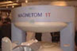
The Dutch vendor chose the organization's sixth annual conference to unveil its work-in-progress Gyroscan Intera 3-tesla MRI scanner, currently being developed in collaboration with the University of Zurich in Switzerland. Philips believes the system will assist brain-mapping efforts through enhanced standard imaging, MR spectroscopy, diffusion, perfusion, and functional MRI (fMRI) techniques.
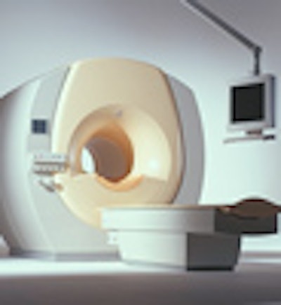
The size of the scanner's patient opening will be the same as in all Intera products, and the scanner will offer all of the Intera line's imaging capabilities as well, including ultrafast image reconstruction with Philips' RapidView reconstructor, high gradient performance, and interactive scanning, according to the firm.
Philips plans to apply for Food and Drug Administration 510(k) clearance within the year, with commercial availability targeted for the end of 2001, according to a company spokesperson. The 3-tesla scanner will be priced in the $3 million range, depending on options and accessories.
In other Philips MRI news, the company also used the HBM show to highlight its SyncraScan package, including the work-in-progress SENSE ultrafast technique, also being developed in collaboration with the University of Zurich. Philips claims that SENSE will double MRI scanning speed.
Philips is also featuring a number of fMRI enhancements, including its work-in-progress real-time fMRI technology. By allowing for real-time image acquisition, reconstruction, and analysis, Philips believes real-time fMRI will allow users to end image acquisition as soon as they are confident that the fMRI exam is successful.
Another product, diffusion tensor imaging (DTI), allows for viewing of the brain's information highway, according to Philips. DTI works by taking raw diffusion-weighted imaging (DWI) data and subjecting it to a computerized mathematical calculation called a tensor field, according to Philips.
The end products of DTI are maps by which researchers can discern individual fiber tracts and determine their direction and course, from their origin in the white matter of the brain to their connections in the cortical gray matter, according to Philips. Philips is collaborating on the development of DTI with the F.M. Kirby Research Center for Functional Brain Imaging at the Kennedy Krieger Institute in Baltimore.
Finally, Philips pointed to its 3D Presto technique, which the vendor believes improves brain perfusion imaging. With the technique, critical blood flow parameters such as relative cerebral blood volume (rCBV), time-to-peak (TTP), and mean transit time (MTT) can be measured, yielding calculated maps that could be used for qualitative assessment of perfusion changes in tumors, stroke, or neurodegenerative diseases, according to Philips.
Philips claims that 3D Presto -- which features an acquisition speed of less than two seconds -- is the only technique currently available for generating whole-brain, 3-D information on cerebral perfusion.
AuntMinnie.com staff writer
June 15, 2000
Related Reading
Philips enters Open MRI with launch of Panorama at ECR, March 7, 2000.
Let AuntMinnie.com know what you think about this story.
Copyright © 2000 AuntMinnie.com