Canadian scientists are presenting research on a new technology that places an MRI scanner onboard a medical linear accelerator at this week's American Association of Physicists in Medicine (AAPM) meeting in Anaheim, CA. The objective of the prototype system is to provide more accurate radiation therapy for difficult-to-treat cancer tumors.
B. Gino Fallone, Ph.D., director of the University of Alberta's Department of Medical Physics at Cross Cancer Institute in Edmonton, is scheduled to discuss the experimental hybrid linac-MRI system in a Tuesday presentation at the AAPM conference.
The hybrid linac-MR system is designed to provide real-time imaging, real-time tumor tracking, and image-guided adaptive radiation therapy simultaneously. Its use is intended to improve the accuracy of radiation treatments for solid tumors and to be able to provide precise radiation therapy treatment for moving tumor sites located in the lung, liver, stomach, prostate, and pancreas.
 |
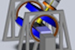
| Schematic of linac-MR system prototype built consisting of the magnet structure (in blue) and associated linac and gantry. X-ray beam, magnet structure, and gradient coils rotate in unison within transverse plane about a single rotational axis. The linac target is on the magnet side of the red cylinder by the item called "linac." All indicated measurements are in mm. Distance from linac target to magnet isocenter is 800 mm. The magnet pole diameter is 632 mm. All images courtesy of Cross Cancer Institute. |
The system's developers also hope that it will minimize the need to radiate a margin of healthy tissue, a practice that is currently required to ensure that the entire tumor has been treated. In addition, its precision will enable a higher dose of radiation to be delivered to cancerous tumors. The technology has the potential to increase the likelihood of tumor control by an estimated 20% to 40%, according to Fallone.
The system developed by the Cross Cancer Institute's Linac MR Research Group is a 6 MV linac mounted on the open end of a biplanar, low-field (0.2 tesla) permanent MRI magnet, with both the linac and magnet on a single gantry that rotates around the patient. It is intended to image the entire tumor continuously and adjust the radiation beams accordingly.
The permanent magnet's poles (32.3 x 32.3 inches2) are rigidly held apart to give an 11-inch pole-to-pole opening with flat gradients (40 mT/m max) running under a console. The linac components consist of a salvaged magnetron-based decommissioned system (Varian 6 MV 600C, Varian Medical Systems, Palo Alto, CA). The distance from linac target to MR isocenter is 31.5 inches, and Faraday case shielding is used.
The head prototype has a 27-cm gap and the whole body prototype has a 70-cm gap. The head prototype unit is fully operational and produced its first images of a tumor in a head phantom in December 2008. The images (128 x 128 pixels) were obtained in approximately 38 seconds using raw gradient echo sequences. During linac radiation, the MRI images had no geometric distortion but a slightly reduced signal-to-noise ratio, Fallone said.
 |
 |
 |
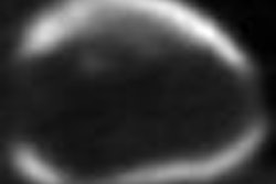
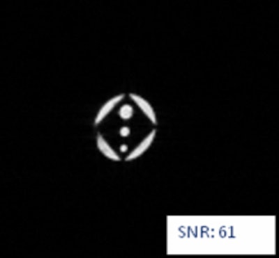
| Top image above, CT of acrylic rectangular cube; second image, MR without linac; third image, MR with linac. Above images, in December 2008, signal-to-noise ratios of MR images differ slightly between images obtained with linac irradiation on and those with linac off. The imaging has improved such that as of May 2009, the signal-to-noise ratios (shown below) are essentially the same. Images below are obtained with thinner slices compared to those above; thus, the reason for different signal-to-noise ratios. |
 |
 |
To overcome the incompatibility issues of an MRI scanner and a linear accelerator working at the same time, the Linac MR Research Group created a specialized design that shields radiofrequency (RF) waves and magnetic fields. Ordinarily, MRI magnets interfere with linac systems, and the radio waves emitted by linac systems to accelerate electrons to high speeds interfere with MRI scanners.
Magnetic shielding prevents the MRI from disturbing the linac, and RF signal shielding, modifications to RF signal triggering, and pulse shaping are used to minimize the linac's electromagnetic interference with the MRI.
The Alberta Cancer Foundation in Edmonton has contributed more than $3 million ($2.8 million U.S.) to the project since it was initiated in 2005. Funding is also being provided by the Alberta Cancer Board, with headquarters in Edmonton, and Alberta Health Services located in Calgary.
In related research, Amit Sawant, Ph.D., an instructor in the Department of Radiation Oncology at Stanford University in Stanford, CA, presented findings on developing imaging specifications to increase image acquisition by an MRI scanner to up to 10 times faster than current system performance. Sawant and colleagues are developing specifications for a system to accurately image the entire volume of a tumor about three times per second.
Sawant believes the key to implementing the technology may be to lower the magnetic field of the magnetic resonance used to image a tumor. During therapy, images do not need to be of the highest quality to track tumors, according to the Stanford research team. An MRI image only needs to show tumor boundaries.
By Cynthia E. Keen
AuntMinnie.com staff writer
July 28, 2009
Related Reading
Linac-MRI prototype allows real-time tumor tracking during rad therapy, September 14, 2007
Copyright © 2009 AuntMinnie.com