Eleven medical associations have released guidance on the use of PET/CT and SPECT/CT for patients with cardiovascular (CV) infections.
The recommendations could improve patient care, as current clinical tools are often insufficient in complicated cases, noted lead author of the guidance Jamieson Bourque, MD, of the University of Virginia Health System in Charlottesville. The document was published jointly March 11 in the Journal of Nuclear Cardiology, Clinical Infectious Diseases, the Heart Rhythm Journal, and JACC: Cardiovascular Imaging.
“The stakes are high with cardiovascular infection because the incidence is increasing and there is associated high morbidity and mortality,” Bourque said in an Infectious Disease Society of America (IDSA) news release.
Valvular infective endocarditis is a bacterial infection of the heart valves, leading to growths that can disrupt normal function and that can lead to heart failure and stroke. In the U.S. and elsewhere, studies suggest the incidence of these infections is increasing due to higher rates of device implants and the recent epidemic of injection drug use, according to the authors.
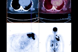
In PET/CT and SPECT/CT exams, patients are first injected with radiotracers that target the site of the infections, with the scanners then revealing the activity.
In the article, experts provide evidence-based consensus on specific clinical scenarios where PET/CT and SPECT/CT add value for patient care. The recommendations emphasize the complementary nature of these advanced nuclear medicine exams.
“Current strategies based on clinical criteria and an initial echocardiographic imaging approach are effective but often insufficient in complicated cardiovascular infection,” the authors wrote.
Specifically, the recommendations outline indications for echocardiography, cardiac CT angiography, radiolabeled leukocyte SPECT/CT, and F-18 FDG-PET/CT for assessing patients. The authors provide clinical indication ratings of “Appropriate,” “May Be Appropriate,” or “Rarely Appropriate” for use of the tests in 73 clinical scenarios.
In addition, the recommendations include the following:
- Diagnostic algorithmic flowcharts for suspected native or prosthetic valve infective endocarditis or prosthetic material/ventricular assist device (VAD) infection and for suspected cardiovascular implantable electronic device infection (CIED) infection
- Teaching images from cases in which F-18 FDG-PET/CT and SPECT/CT studies were used in prosthetic valve endocarditis, CIED pocket and lead infection, VAD infection, and prosthetic material infection
- Teaching case examples in which F-18 FDG-PET/CT and SPECT/CT were used to assess prosthetic valve endocarditis, suspected lead CIED infection, suspected VAD infection, and suspected prosthetic material infection
“This advanced imaging can aid in key medical and surgical considerations,” the group wrote.
Ultimately, the authors noted that the process of developing the recommendations identified gaps in the literature requiring future research, with such investigations expected within the next few years to provide additional revisions.
“We intend for this consensus statement to be used as a model for inclusion of nuclear imaging into a multimodality approach with feedback incorporated from relevant clinical and imaging societies,” the group concluded.
The full recommendations can be found here.