Brain PET imaging with carbon-11 methionine (C-11 MET) may be an effective new approach for identifying developmental brain tumors in children and adolescents, according to a study published February 7 in PLOS One.
A South Korean group looked at imaging findings in patients with intracranial germinomas who underwent C-11 methionine (MET) PET/CT scans and compared results with standard clinical lab variables for diagnosing the tumors. The team found that C-11 MET performed as well as clinical lab variables and could serve as a tool to help clinicians find the tumors before they potentially spread.
"This modality may have a diagnostic role in patients suspected of having [intracranial germinomas], especially for localizing tumors," wrote principal investigator Dr. Seung Hwan Moon of Sungkyunkwan University School of Medicine in Seoul.
Germinoma is a rare type of tumor and is most often found in the brains of children between the ages of 10 and 19. The tumors arise from germ cells, which normally become eggs or sperm after migrating to the ovaries or testes during fetal development. When these germ cells fail to migrate to proper locations, however, they may continue dividing and form tumors.
Of two types of germinomas, intracranial germinomas are typically slower growing and respond well to treatments such as chemotherapy, while intracranial nongerminomas secrete chemicals into the spinal fluid and bloodstream and require more intensive treatment.
Early diagnosis of these tumors with conventional brain MRI can be difficult due to their slow progression or subtle signal changes, according to the authors. C-11 MET-PET/CT is a proven imaging tool in brain tumors, yet due to the rarity of intracranial germinomas, its value in diagnosing the disease has not yet been fully investigated, the authors wrote.
In this retrospective study, the group analyzed results from 21 consecutive patients (average age, 16 years old) with confirmed intracranial germinomas and eight patients with intracranial nongerminomas who underwent C-11 MET-PET/CT scans (Discovery STE, GE Healthcare) at Samsung Medical Center from December 2013 to May 2020.
Clinical characteristics, imaging findings, and tumor markers such as α-fetoprotein (AFP) and beta-human chorionic gonadotropin (HCG) in serum and cerebrospinal fluid were used as clinical variables. Maximum standardized uptake value (SUVmax), tumor-to-normal tissue (T/N) ratio, and visual scoring of tumor were used as C-11 MET-PET/CT parameters.
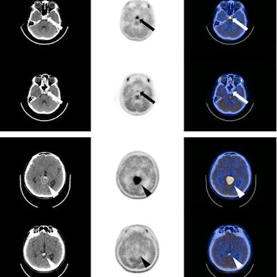
 Noncontrast CT, MET-PET, and fusion images of two representative cases in patients with intracranial germinomas before and after treatment. (A, B) The first case was an 11-year-old boy who experienced headache, polydipsia, polyuria, and growth hormone deficiency before treatment. The primary tumor was located in the sellar-suprasellar region (white and black arrows). Before treatment, pre-Tx SUVmax and pre-Tx T/N ratio were 4.91 and 4.38. After chemotherapy and radiation therapy, post-Tx SUVmax and post-Tx T/N ratio decreased to 1.62 and 1.27. The visual grade was 3 before the treatment and decreased to 2 after treatment. (C, D) The second case was a 14-year-old boy who experienced headache before treatment. The primary tumor was located in the pineal region (white and black arrowheads). Before treatment, pre-Tx SUVmax and pre-Tx T/N ratio were 4.95 and 3.96. After chemotherapy and radiation therapy, post-Tx SUVmax and post-Tx T/N ratio decreased to 1.52 and 1.15. The visual grade was 3 before treatment and then decreased to 1 after treatment. Image courtesy of PLOS One.
All of the intracranial germinomas were well visualized on C-11 MET-PET/CT and the SUVmax of intracranial germinomas was higher than that of intracranial nongerminomas (p = 0.005), according to the findings.
In addition, the authors identified significant correlations between PET parameters and clinical HCG levels. T/N ratio was significantly correlated with serum HCG (p = 0.014) and cerebral spinal fluid HCG (p = 0.010), while SUVmax of C-11 MET also showed a significant correlation with cerebral spinal fluid HCG (p = 0.048).
The authors noted limitations, namely that this was a single-center study with a small number of patients and a retrospective design. The results should be verified in a future multicenter study involving a large number of patients with a prospective design, they wrote.
Nonetheless, C-11 MET-PET/CT appears to have diagnostic value in patients with intracranial germinomas, with C-11 MET avidity as a potential surrogate biomarker of HCG, a standard clinical diagnostic marker for the tumors, they wrote.
"Some difficult cases of [intracranial germinomas] cannot be diagnosed by conventional MRI; in these cases, PET imaging could be considered," the authors concluded.