Though whole-body MRI is highly accurate in detecting soft-tissue abnormalities, the modality falls short in finding metaphyseal lesions and rib fractures in cases of suspected child abuse, according to a study in the September issue of the American Journal of Roentgenology.
Researchers at Children's Hospital Boston and Harvard Medical School in Boston concluded that whole-body MRI "was inferior to radiography in the detection of rib fractures and classic metaphyseal lesions, which are high-specificity indicators of infant abuse that constitute 90% of noncranial skeletal injuries in infant abuse fatalities."
The lead author of the study was Jeannette Perez-Rossello, MD, a pediatric radiologist at Children's Hospital Boston (AJR, September 2010, Vol. 195:3, pp. 744-750).
Perez-Rossello and colleagues also noted that whole-body MRI had high specificity but low sensitivity for identifying fractures, compared with combined information from initial and follow-up skeletal surveys.
Patient population
The retrospective study reviewed imaging findings for 21 infants, ranging in age from 0 to 12 months (mean age, 5.4 months), who received a whole-body MRI scan between March 2003 and October 2005 to evaluate skeletal and soft-tissue injuries.
Among the injuries that raised concerns of possible physical abuse were subdural hemorrhages, cerebral edema, retinal hemorrhages, and highly specific fractures, such as classic metaphyseal lesions, rib fractures, multiple fractures, and different stages of healing.
Among the 21 infants in the study, a child protection team concluded that all but one child had been physically abused. The exception was an infant initially thought to have been abused, but who instead was diagnosed with osteogenesis imperfecta.
Whole-body MRI exams were conducted on a 1.5-tesla system (Signa, GE Healthcare, Chalfont St. Giles, U.K.), with patients imaged with a phased-array coil and a maximum field-of-view of 48 cm for wide body coverage from the shoulders to the heels.
The skeletal survey images were acquired with a high-detail film-screen combination (13 line pairs/mm, 26 studies) or a high-detail dual-side computed radiography system.
Image evaluation
Two board-certified pediatric radiologists compared imaging results and agreement between the initial skeletal survey and the follow-up skeletal survey, the initial skeletal survey and the whole-body MRI, and the summary skeletal survey and the whole-body MRI.
The researchers found a total of 167 areas of fracture or signal abnormality. The summary skeletal survey identified 114 (68%) fractures or areas of signal abnormality, compared with 99 (59%) by whole-body MRI and 95 (57%) by the initial skeletal survey. In addition, 68 (41%) fractures or areas of signal abnormality were seen only on summary skeletal survey, compared with 53 (32%) seen only by whole-body MRI.
There was no statistically significant difference between the average number of fractures or signal abnormalities detected by whole-body MRI and radiography per patient. Whole-body MRI identified an average of 4.7 areas of signal abnormality, while the summary skeletal survey identified an average of 5.4 fractures.
Metaphyseal lesions
The study also identified a total of 37 classic metaphyseal lesions or metaphyseal hyperintense areas. Twenty-four (65%) were seen by summary skeletal survey only, two (5%) were seen by whole-body MRI only, and 11 (30%) were seen by both techniques.
"Agreement between the classic metaphyseal lesions seen in the summary skeletal survey and the whole-body MRI was poor," the authors wrote, with a corresponding concordance rate of 29%.
Whole-body MRI identified 11 of 35 classic metaphyseal lesions, which were seen in the summary skeletal survey. Whole-body MRI also registered a low sensitivity of 31% in identifying signal abnormalities in areas where classic metaphyseal lesions were detected by radiography.
Rib fractures
In the area of rib fractures, radiography outperformed whole-body MRI. The fracture location and the presence of healing could be determined with the skeletal survey but was not possible with whole-body MRI.
The summary skeletal survey identified 33 regions of one or more rib fractures, while whole-body MRI identified 32 regions of signal abnormality in the rib cage. There was low agreement between the regions of hyperintensity seen with whole-body MRI and the regions of rib fractures identified in the summary skeletal survey. The corresponding concordance rate was 41%.
 |
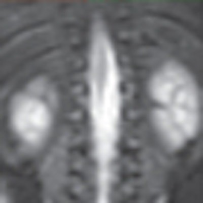
| A 3-month-old abused boy was brought to the emergency department with facial bruises and multiple fractures. Initial anteroposterior radiograph (A) of the chest shows multiple posterior rib fractures (arrows). Coronal MRI short-tau inversion recovery (STIR) image (B) of the chest shows no signal abnormality in posterior ribs or chest wall. Images courtesy of the American Journal of Roentgenology. |
Whole-body MRI had low sensitivity of 57% in identifying signal abnormality in areas of rib fractures seen with radiography.
"Although whole-body MRI performed somewhat better for the detection of rib fractures, with moderate agreement with the skeletal survey reference standard, sensitivity of 57% is discouraging," the authors noted. "This limitation is further compounded by the inability of whole-body MRI to localize injuries to specific ribs."
Whole-body MRI, however, was able to detect additional findings in soft tissue. In some cases, the discovery of soft-tissue hyperintensity by whole-body MRI helped to identify a fracture not seen in the skeletal survey, but which was seen healing on the follow-up study.
Based on the results, the researchers concluded that while there was no significant difference between the radiographic skeletal survey and whole-body MRI in the total number of skeletal injuries identified, whole-body MRI "was inferior to radiography in the detection of rib fractures and classic metaphyseal lesions."
In addition, there was only "fair agreement between the classic metaphyseal lesions identified by the summary skeletal survey versus metaphyseal signal abnormalities on whole-body MRI," the authors noted. "The failure of whole-body MRI to detect even one in three classic metaphyseal lesions identified on the skeletal survey precludes its use as a substitute for radiography in the initial imaging assessment of suspected infant abuse."
By Wayne Forrest
AuntMinnie.com staff writer
August 24, 2010
Related Reading
High-field MRI appropriate for fetal autopsy, August 18, 2009
MRI with STIR fails to change diagnosis in child abuse cases, May 12, 2009
MRI helps find fractures among elderly female ED patients, April 29, 2009
MRI used to diagnose complex lung infections in children, October 16, 2008
MR parameters divulge depth of acute hamstring injuries, July 13, 2007
Copyright © 2010 AuntMinnie.com