Portable, wireless dual-energy imaging may soon become a valuable resource for bedside applications: Researchers have had early success with a prototype system that has greatly enhanced the view of interventional devices implanted in phantoms and cadavers.
A team of biomedical engineers from Johns Hopkins University (JHU) in Baltimore and Carestream Health in Rochester, NY, are collaborating on the technology, which has produced better image quality than previous dual-energy technology, with no more radiation dose than an average radiograph.
"We now know the high and low energies we should be delivering and we know the doses we should be delivering so the total dose is equivalent to a conventional x-ray," said Jeff Siewerdsen, PhD, associate professor of biomedical engineering at JHU. "And we know the algorithms for decomposing a dual-energy image that make something like a tube or needle pop right out of the image."
Siewerdsen presented details of the project at the recent American Association of Physicists in Medicine (AAPM) annual meeting in Philadelphia.
'Underappreciated technique'
''Dual-energy imaging is an underappreciated technique," Siewerdsen said. "Some early entries into the clinical use of dual-energy imaging have been suboptimal. It has been either too slow, image quality has been too poor, or it has involved increasing radiation dose."
Despite those shortcomings, dual-energy imaging has shown that it can distinguish between specific materials in the body. "The classic example is its ability to knock out calcium from an image, which leaves an image of soft tissue, or to knock out the soft tissue and leave just an image of calcium," he added. "In the same way, we can knock out materials and leave the image of just the interventional device, because its material characteristics are very different from its surroundings."
JHU and Carestream began working on the dual-energy imaging technology about five years ago. The partners developed a dual-energy radiography system for chest imaging and conducted a clinical trial with 220 patients. That dual-energy system was able to enhance the detection of lung nodules in a comparison with digital radiographs at the same radiation dose.
Portable applications
With that knowledge in hand, Siewerdsen and colleagues turned their attention to portable imaging applications. Although bedside and portable imaging inside an intensive care unit may be the most common radiographic exam performed, Siewerdsen believes it also probably has the worst image quality of any modality.
"The reasons include difficulty in setting up a film-screen cassette or computed radiography cassette behind the patient in bed and aligning the x-ray source on the film cassette, which is a geometric challenge," he added. "In terms of image quality, the major factors are quality of the detector and scatter, which is a giant problem in portable radiography."
 |
| JHU and Carestream used this experimental configuration for its dual-energy system to image a phantom. All images courtesy of Jeff Siewerdsen, PhD, Johns Hopkins University. |
Technical challenges
Before the system, which features Carestream's DRX-1 digital radiography (DR) wireless flat-panel detector, can be considered for human trials, there are two technical challenges to conquer. The first hurdle is that the system must deliver high-energy and low-energy beams within one second of each other to minimize motion artifact. Similarly, the DR detector must be able to read the two images in less than one second.
To enhance the system's ability to read images faster, researchers are working on two approaches. The first step is to add memory to the detector. "DR detectors are intrinsically very fast and this one is," Siewerdsen said. "You need to be able to read two images in a row, store them to memory on the detector, and read them all through this wireless connection." That solution, he said, is within reach.
The second approach is using the DR detector for real-time readouts, rather than as double-reads, like other DR detectors are used in fluoroscopy.
"It's not that we need another year to overcome some technical hurdle; there is no technical hurdle," Siewerdsen said. "All that is required is the will to implement fast readout on a portable detector. The onus is as much on people like me to show that this is something we really need in the intensive care unit to improve image quality at the bedside, reduce the number of retakes, and improve patient safety, because we are getting good reads fast at the bedside."
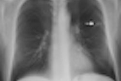
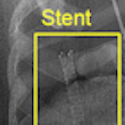
 |
| Dual-energy imaging shows the placement of a stent in a cadaver more clearly than the stent can be seen by digital radiography. |
Researchers also expect this dual-energy technology to have the same low dose of radiation as a common radiograph. "Our initial objective was to build a dual-energy system that could increase performance without increasing the dose above a radiograph," Siewerdsen said. "If we achieve that, I think we should go further. Even if the dose of a radiograph is low, if we can get an image that accomplishes the task at a lower dose, we should."
Siewerdsen and colleagues currently are planning to finish technical development of the system, which means optimizing the dual-energy imaging technique and image processing. The next step will be to connect the system to a faster DR detector before moving to patient trials.
By Wayne Forrest
AuntMinnie.com staff writer
August 18, 2010
Related Reading
Researchers eye dual-energy DR as calcium-scoring option, May 5, 2010
Will new DR technologies offer alternative to CT? Part 1, March 24, 2010
Virtual dual-energy DR improves lung lesion detection, February 10, 2010
Rads prefer dual-energy x-ray over standard DR in lung, January 29, 2009
Dual-energy digital x-ray still looking for acceptance, February 22, 2007
Copyright © 2010 AuntMinnie.com