A Florida company is raising eyebrows in marketing a fluoroscopy system to chiropractors via a website that touts the unit's ability to generate higher insurance settlements in cases of chronic spinal trauma. The issue could be another front in the ongoing debate over the appropriate clinical use of medical radiation.
DMX Works of Palm Harbor is selling its Digital Motion X-ray (DMX) low-dose C-arm fluoroscopy system to chiropractic offices for $90,000. The company claims the system can reveal signs of soft-tissue injuries based on dynamic images that can be missed with more advanced modalities such as MRI and CT.
Critics, however, are concerned about the radiation dose delivered by the system, and whether the procedures are medically indicated.
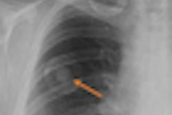
On its website, DMX Works touts the DMX system as a "new type of fluoro-based x-ray system" that uses "new digital and optic technology" to enable clinicians to view the spine in real-time motion at 30 frames per second. The company claims that the system offers a superior method for diagnosing whiplash injuries compared to static modalities such as CT and MRI, because whiplash patients "hurt more when they move."
But some critics have doubts about the clinical value of the technology, and they also question the appropriateness of the company's sales pitch to chiropractors: "DMX can dramatically increase settlement amounts for people who have sustained ligamentous injuries that MRI, CT, and static x-rays cannot detect," the website claims, citing several million-dollar settlements that were obtained by using the system.
Representatives from DMX Works did not respond to calls and e-mails for comment.
Clinically indicated?
In a world in which highly advanced technologies such as MRI and CT are available, is there still a role in diagnosing ligament injuries for video fluoroscopy, which was first developed in 1896? It may depend on who you ask.
Mitchel Harris, MD, a Boston orthopedic surgeon, noted that orthopedic physicians and chiropractors use different terms to describe ligament injuries, and pointed out that fluoroscopy motion studies can't show ligaments.
"A chiropractor may say a patient's back is unstable, but his definition isn't the same as mine. This technique may show motion of the spine that is more than normal but might not qualify as instability by orthopedic or spine standards," he told AuntMinnie.com. "One technique is dynamic; one is static. An MRI shows structural injury that will relate to instability; motion studies will accentuate that instability."
But he acknowledged that fluoroscopy units can show "hypermobility."
Both modalities can show ligament injury, but fluoroscopy shows the motion of bones because it's done in a dynamic fashion, which MRI cannot do, he noted.
Harris questioned the validity of some the company's claims of the system's superior ability to detect ligament injuries, but said that doesn't mean they are completely specious, especially for patients who suffer chronic pain after a whiplash accident.
"[Fluoroscopy] can infer what ligaments are doing," Harris noted. "They can be stable and don't need surgery, but it doesn't mean it doesn't hurt. If they're doing it for a bigger settlement because it hurts more and tests show it's moving, it's not necessarily a false claim."
Phillip Tirman, MD, a radiologist and musculoskeletal MRI director for the Northern California division of imaging services firm RadNet, said that focusing on instability opens up a clinical gray area.
"It is the secondary effect of ligament damage, not visualization of the ligament damage itself, which they are claiming to see that MRI can't," he said in an e-mail to AuntMinnie.com. "MRI directly visualizes the torn ligament; this machine doesn't. Also, making the diagnosis of ligament damage due to perceived instability is very subjective, and there will be a lot of unscrupulous individuals who would use this subjectivity for monetary gain."
Beneficial if used properly?
But chiropractic radiologist Glynna Rangel, DC, a diplomate of the American Chiropractic Association's board of radiology and vice president of the group's Council on Diagnostic Imaging, told AuntMinnie.com that the "modality is definitely beneficial if used properly, especially in the upper cervical spine."
"Video fluoroscopy will show a tear in the ligament, which causes instability and demonstrates motion that would occur due to ligament injury that you can't see on MRI," she explained. "MRI, which is primarily used for pathology, shows acute ligament injury but not functionality."
Rangel noted that she has seen a few cases where MRI didn't reveal ligamentous problems because the injury was old and there was no inflammation or hemorrhage in the area. "[The patient] ended up having to have surgery because of what was shown on the video fluoroscopy study," she pointed out.
However, Rangel, too, takes issue with the company's claims. "They take it a little too far as far as marketing," she said. "Video fluoroscopy is of some value, but I think they're extending it a little bit further than it truly is. They say they can diagnose a little more than they really can, and it doesn't take into effect other variables."
For example, she said that if a patient rotates slightly during the video fluoroscopy, the movement will produce a different image. "Something I would call normal motion, they would say is abnormal; they might say the motion shows injury to this ligament, and it's not always that way," Rangel said. "They take it a step too far and don't incorporate the whole picture."
Rangel said she also has a problem with the company's claims of enhanced insurance settlements. "It completely defeats the purpose of video fluoroscopy, which has specific protocols which should be followed before they even use it, and I found that's not always so," she observed.
The American Chiropractic Association has expressed concern about the inappropriate use of video fluoroscopy in its guidelines.
But Ed Harkins, a Long Beach, CA, chiropractor who bought a DMX system in 2004, said it provides "a piece of the puzzle" in determining ligamentous injuries. He estimated that he uses the video fluoroscopy system on 20 patients per month; most of the cases involve car accidents, workers' compensation claims, or sports injuries.
"It demonstrates injuries that have been missed on other modalities like MRI, CT, and plain x-ray films," he told AuntMinnie.com. "It's able to find a path in the upper spine, alar, and accessory ligaments, which cause upper neck pain after car accidents. All MRI focuses on is the C2 vertebrae and below."
Garry Gold, MD, an associate professor of radiology at Stanford University in Stanford, CA, said orthopedic surgeons commonly do fluoroscopy studies, but they are done during surgery.
"I think their claims are poor," Gold told AuntMinnie.com by e-mail after reviewing DMX's website. "X-ray cannot see soft-tissue structures, and ligament and other soft-tissue damage is very accurately diagnosed by MRI. They may be able to detect abnormalities by abnormal shifting of the bones, say with flexion and extension of the spine, which is something we see routinely as radiologists anyway on static flexion and extension views."
James Taylor, a Kentucky radiologic technologist, rented the mobile version (the unit is installed in a van) of the DMX system for a few months to check out its business prospects. "They wanted us to rent it from them and market it to chiropractors," he said.
He took the system to long-term care facilities to film swallow studies and to chiropractors' offices to do neck studies.
"[Chiropractors] charged ungodly amounts of money, like $2,000 per hour, for studies," he told AuntMinnie.com. "It just didn't seem right; the patients' symptoms weren't there."
Radiation dose
One of the hottest issues in radiology is radiation dose, with regulators and clinicians looking for ways to reduce unnecessary procedures and to reduce the dose delivered when imaging studies are clinically indicated.
How much radiation does the DMX system deliver during procedures? Several contacts for this story gave varying estimates: Industry experts say a typical 15-sec film probably delivers about 20 mSv, but California chiropractor Harkins, who uses the DMX system, says it delivers much less radiation than a chest x-ray or CT scan.
According to the company's website, the system delivers "2,700 x-ray images with the same amount of radiation as a standard x-ray machine, which gives you 6 static x-rays."
"They always marketed it as having less radiation than a regular x-ray machine doing a spinal series with flex and extensions," Taylor said. "But common sense tells you if you're taking thousands of x-rays per minute, fluoroscopy puts out more radiation because it's constantly on compared to a conventional x-ray machine."
But chiropractic radiologist Rangel disputed that, saying the system's image intensifier is designed to reduce the amount of radiation in fluoroscopy studies because they take longer. "A cervical spine video is equivalent to a cervical Davis series with regular x-rays," she said.
The units also featured microphones which, Taylor claims, are designed to record bone popping. "It was all to impress attorneys -- to dramatize the injuries for the courts," he said.
"The system may have some value but not in the application they're using it for," Taylor concluded. "The whole thing just smelled really bad."
By Donna Domino
AuntMinnie.com contributing writer
June 23, 2010
Copyright © 2010 AuntMinnie.com