New techniques in synchrotron x-ray imaging have given researchers a surprising glimpse into our collective past: An international group has obtained detailed information about the brain structure of Australopithecus sediba, a new species thought by some to be the true ancestor to modern humans.
Paleontologists have frequently used medical imaging modalities such as CT to gain insight into archeological specimens without harming them. But South African researchers had to resort to an even more powerful technology -- synchrotron microtomography -- after medical CT failed to provide the required level of detail. In the process, they've discovered key findings about the brain composition of Australopithecus sediba that may bring about changes in evolutionary theories.
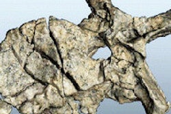
 3D rendering of the skull of Australopithecus sediba from x-ray data gathered at the ESRF. Image courtesy of ESRF and Paul Tafforeau, PhD.
Specifically, while the fossil's brain showed indications of neural reorganization resembling that found in modern humans, the volume, at 420 cm3, was much smaller than expected, countering the theory that the human brain gradually increased in size as it evolved structurally, according to Kristian Carlson, PhD, from the University of the Witwatersrand in South Africa, and colleagues (Science, September 9, 2011, Vol. 333:6048, pp. 1402-1407).
The discovery was brought about by the use of propagation phase-contrast x-ray synchrotron microtomography to image the skull, the "gold standard for imaging fossils since it is the most powerful, nondestructive 3D imaging technique presently available," the authors wrote.
Yet synchrotron scanning also posed a challenge, according to the group, in that a new protocol needed to be used to image a fossil of that size -- and one still filled with the rock in which it was buried.
A new species
The current study is part of a collection of five papers published in the journal this month that further characterize Australopithecus sediba, providing information on the pelvis, hand, foot, and ankle, in addition to the endocast, or cranial cavity, of the species.
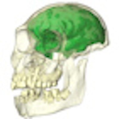
 A reconstruction of the juvenile skull (partially transparent) with the brain endocast depicted in green. Image courtesy of Witwatersrand University and Kristian Carlson, PhD.
"The very many advanced features found in the brain and body make it possibly the best candidate ancestor for our genus, the genus Homo, more so than previous discoveries such as Homo habilis," said co-study author Lee Berger, PhD, also from the University of the Witwatersrand, in a statement on the research.
It was actually Berger's young son, Matthew Berger, who discovered the first specimen -- a right clavicle -- of the two partial skeletons that were buried in cave deposits at a site in Gauteng, South Africa (Science, April 9, 2010, Vol. 328, pp. 195-204).
The skeletons of a juvenile, with brain growth essentially complete, and an adult are believed to be 1.95 million to 1.78 million years old. "Together they represent a new species of Australopithecus that is probably descended from Australopithecus africanus," Berger and colleagues wrote.
More powerful imaging
By examining the inner surface of the skull, the researchers hoped to recreate the shape and size of the brain. While the skull of the juvenile was originally examined with a medical CT scanner, the data did not allow detailed investigation of the internal structures, according to Carlson and colleagues.
To obtain more information, the researchers decided to examine the skull at the European Synchrotron Radiation Facility (ESRF) in Grenoble, France, where paleontologist and co-author Paul Tafforeau, PhD, had been studying synchrotron imaging for fossil primate teeth.
"The original aim was to study the teeth, as it is my research field; at that time, the skull was not yet discovered," Tafforeau told AuntMinnie.com. "After the discovery of the skull, we designed the synchrotron experiment for complete imaging of the skull. We went for the synchrotron approach because no other technique would have been able to produce such high-quality data on a fossil skull that is still partially embedded in a strong rock matrix."
X-ray synchrotron microtomography uses radiation produced by a circular particle accelerator, or synchrotron. In a synchrotron, particles subject to electric and magnetic fields are directed through a large storage ring. As the beam moves through the storage ring, specific magnet configurations (e.g., "wigglers") deflect the particles, creating much stronger x-rays than with other types of systems.
The radiation produced from the beam is captured by straight beamlines that branch off of the ring. Fossils or other objects can be placed at the ends of these beamlines for imaging. Using a beamline at the ESRF, the group was able to scan the skull at a resolution (3D pixel size) of approximately 45 microns.
 A comparison of the juvenile skull imaged with medical CT (left) versus synchrotron scanning (right). Voxel size is approximately 450 µm for the medical CT scan, compared with 45.71 µm with synchrotron imaging. Image courtesy of ESRF and Paul Tafforeau, PhD.
Blocked by rock
"Imaging a partial hominum cranium filled with matrix and still achieving a resolution exceeding 100 µm (i.e., voxel size smaller than 50 µm) with phase contrast presented a considerable challenge," Carlson and colleagues wrote. Previously, synchrotron imaging had generally been limited to smaller fossils.
One difficulty is that the larger size of the stone-filled cranium (approximately 15 cm) resulted in too low of a transmission level, or the ratio between the direct beam and the beam after passing through the sample, according to the authors. Even with the strongest energy level available (150 keV), with a direct beam at the limit of the camera's saturation, high-quality images could not be obtained.
To address this, they used a protocol that "substantially increases the amount of photons going through the middle of the sample, but without reaching the saturation level of the detector," Carlson and colleagues wrote. By adding a configuration of aluminum in front of the beam, as well as placing the cranium within a specially designed Plexiglas cylinder filled with aluminum or glass balls, they were able to effectively "normalize" x-ray absorption across the entire field-of-view.
The protocol continues to be tweaked as it's used for other dense or difficult specimens, Tafforeau said.
"I still continue to improve it," he said. "For the moment, we use glass balls or aluminum balls, but I would tend toward more amorphic systems and to push the concept to even submicron resolution. Nevertheless, finding the adequate substance to control absorption at submicron resolution with very high x-ray dose, without danger for the fossils, is not an easy task."
While keeping the rock inside of the skull added challenges to the imaging process, keeping everything intact helped preserve the inner surface.
"It makes the scan more difficult," Tafforeau noted, "and also the segmentation had to be done manually. Nevertheless, as physical preparation can make fossils fragile, we decided to go for a virtual preparation. In a way, it is a solution to ensure good preservation of this fossil for the next generations of paleoanthropologists."
Surprising findings
These future generations will now have a new theory to investigate, due to the study's surprising results. Carlson and colleagues had expected the size of the brain to be larger, fitting the idea that the human brain increased gradually in size as cortical reorganization occurred.
However, after comparing various measurements of Australopithecus sediba to those from previously discovered fossils, they found a surprising mix of characteristics: While the fossil's overall brain shape resembled that of humans more than chimpanzees, its 420 cm3 volume is on average only about 40 cm3 larger than that of chimpanzees.
"The relative importance and timing of two critical processes in the evolution of the human brain -- cortical reorganization and size increase -- has been debated since the discovery of Australopithecus," the authors wrote.
"Indeed, one of our major discoveries is that the shape and form of sediba's brain is not consistent with a model of gradual brain enlargement, which has been hypothesized previously for the transition from Australopithecus to Homo," Carlson said.