Given its "excellent correlation" to colonoscopy and pathology results, researchers from Rhode Island are recommending that MR enterography be considered as a first-line test for Crohn's disease to reduce radiation from CT scans.
The study, from Warren Alpert Medical School at Brown University in Providence, also indicates that readers are confident in their diagnoses of Crohn's disease, despite reduced image quality of MR enterography compared to CT.
"CT enterography is considered the gold standard imaging modality for the detection and evaluation of Crohn's disease, but it delivers up to five times the radiation dose of small-bowel follow-through, which essentially is the test that [CT] replaced," said lead study author David Grand, MD, director of body MRI and assistant professor of diagnostic imaging. "Patients with Crohn's disease receive higher levels of radiation than the general population. This has been shown in multiple studies and is due predominantly to repeated exposure to CT."
MR versus CT enterography
Grand and colleagues investigated the efficacy of MR enterography for Crohn's disease in two studies. The first study compared back-to-back CT enterography and MR enterography scans performed without antiperistalticagents to restrict bowel contractions.
Patients in this study were referred for CT enterography for known or suspected Crohn's disease. The study excluded patients older than 65 years of age and those with diabetes, known renal dysfunction, and pregnancy. "These exclusion criteria were designed because we had to administer gadolinium," which has been connected to nephrogenic systemic fibrosis, Grand noted during the presentation of results at the 2010 annual RSNA meeting.
All 26 patients received a CT enterography exam immediately followed by an MR enterography scan, generally within 15 minutes. One patient did not complete the imaging cycle because of claustrophobia in the MRI scanner.
The final study population consisted of 13 women and 12 men, with a mean age of 42 years, ranging from 18 to 64 years. Twelve of the 25 patients at the time of the CT and MRI scans were confirmed for Crohn's disease, while 13 patients had no signs of the condition.
The CT protocol included 900 cc of VoLumen (Bracco Diagnostics) beginning 45 minutes prior to scanning and 450 cc of water immediately before imaging on a 16-slice CT system (LightSpeed, GE Healthcare, Chalfont St. Giles, U.K.). Patients also received 130 cc of contrast agent (Omnipaque, GE Healthcare). Axial and coronal images were obtained at 5-mm thickness.
MR enterography scans were conducted on a 1.5-tesla MRI system (Excite, GE Healthcare) with an eight-channel torso-array coil. Intravenous contrast (Magnevist, Bayer HealthCare Pharmaceuticals, Wayne, NJ) was administered at 0.1 mmol/kg for each patient. Axial and coronal images also were obtained.
Two fellowship-trained abdominal readers with experience in interpreting CT and MR enterography images reviewed separately the results for image quality, diagnostic confidence, and the presence and location of Crohn's disease.
"In terms of exam quality, I don't think it was much of a surprise that both readers consistently ranked MR [enterography] image quality subjectively lower than that of CT [enterography], and this was statistically significant," Grand said. "Interestingly, although the readers agree that the quality of the images on MR were inferior to those on CT, it did not affect their ability to make a confident diagnosis, which was rated 10 out of 10 by both readers."
 |
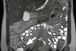
| Images show patient with terminal ileitis. CT (above) and gadolinium-enhanced MR enterography (below) show abnormal wall thickening. Both readers agreed on the interpretation of images. All images courtesy of David Grand, MD. |
 |
MR enterography versus colonoscopy, pathology
The second study was designed to evaluate the sensitivity, specificity, and concordance of MR enterography with colonoscopy and pathology as the gold standards.
Grand and colleagues retrospectively reviewed 850 consecutive MR enterography exams of patients with known or suspected Crohn's disease. MR enterography was conducted on 1.5-tesla systems (Excite, GE Healthcare; Magnetom Symphony and Somatom Esprit, Siemens Healthcare, Malvern, PA) with torso-array and surface coils.
Patients received intravenous contrast at 0.1 mmol/kg (Magnevist, Bayer). Again, no antiperistaltic agents were used.
The researchers compared MR enterography with colonoscopy and pathology reports from all subjects. A total of 310 (36%) of the 850 patients received colonoscopy with a biopsy within 90 days of MR enterography. Of those 310 cases, 162 patients (52%) received a colonoscopy and biopsy within 30 days of MR enterography to ensure the dates of the tests were within a shorter period of time.
Among the 310 patients, colonoscopy with pathology achieved the highest sensitivity (93%), specificity (86%), and positive (86%) and negative predictive values (93%).
"The first thing that is important to note is the concordance between colonoscopy and pathology is not 100%," Grand said. "What the endoscopist sees and what the pathologist sees do not always agree. If you look at the sensitivity and specificity and positive and negative predictive values, they fall short of 100% by a wide margin."
MR enterography compared with colonoscopy achieved sensitivity of 84% and specificity of 76%, while MR enterography compared with pathology reached sensitivity of 71% and specificity of 83%. In addition, results for the 162 patients who received exams within 30 days were quite similar to the results for the 90-day group.
All 310 patients scanned within 90 days
|
162 patients with colonoscopy within 30 days
|
"Interestingly, the numbers for colonoscopy and pathology don't change at all, which is expected because all [the tests] were done on the same day," Grand said. "The MR [enterography] to colonoscopy numbers improved slightly but were not statistically significant. We achieved a sensitivity of 84%, specificity of 82%, and negative predictive value of 86%."
The researchers also evaluated the patients with MR enterography for whom the colonoscopy and pathology were both positive. Grand said that, theoretically, these patients have more severe Crohn's disease because both the endoscopist and pathologist see the condition.
MR enterography compared with both colonoscopy and pathology achieved sensitivity of 83%, specificity of 98%, positive predictive value of 88%, and negative predictive value of 84%.
Grand concluded that MR enterography "shows excellent correlation between colonoscopy and pathology. Antiperistaltic agents may not be necessary, and MR enterography should be considered as a first-line test in this patient population."
By Wayne Forrest
AuntMinnie.com staff writer
February 7, 2011