Adaptive statistical iterative reconstruction (ASIR) of CT data cuts noise and boosts diagnostic confidence of abdominal lesions while slashing radiation dose, reported researchers who used an ASIR algorithm instead of traditional filtered back projection.
The study, published online before print (Radiology, September 9, 2010), was unusual for its acquisition of additional CT views in patients who had already undergone abdominal CT scans. The study was also larger than earlier clinical research, despite its small size.
Researchers from Massachusetts General Hospital in Boston aimed to compare image quality, lesion detection, and lesion conspicuity on abdominal CT images acquired at different x-ray tube current-time products (50-200 mAs).
Images were reconstructed using both filtered back projection (FBP) and the proprietary ASIR algorithm developed by GE Healthcare of Chalfont St. Giles, U.K.
"This algorithm takes into account precise modeling of the x-ray photon statistics and electronic noise, all of which are less accurate in FBP, wrote Sarabjeet Singh, MBBS; Mannudeep Kalra, MD; and colleagues from MGH, along with Jiang Hsieh, PhD, from GE Healthcare.
To cut reconstruction times, "ASIR utilizes the information contained in the FBP-reconstructed image as an initial 'building block' in the reconstruction process," the team wrote. "Reduction of noise and artifacts in low-radiation-dose images are especially helpful in young and obese patients, in clinical situations like multiphase CT examinations, and in patients who have undergone multiple prior examinations."
Singh and colleagues recruited 22 patients (mean age, 60.1 years ± 7.3; range, 52.8-67.4 years; mean weight, 78.9 kg ± 18.3; 12 men, 10 women) who underwent abdominal scans with CT followed by four additional image series for the study.
After injection of 80 to 100 mL of an intravenous contrast medium (iopamidol 370, Bracco Diagnostics, Princeton, NJ), abdominal images were acquired on a 64-detector-row scanner (Discovery CT750 HD, GE Healthcare).
Two experienced radiologists examined the images, recording CT dose index volume (CTDIvol), dose-length product (DLP), patient weight, objective noise, and CT attenuation numbers, analyzing the data using analysis of variance and the Wilcoxon signed-rank tests.
There was some variability in interobserver agreement between the two readers (κ = 0.12-1.0); however, the percentage agreement between the two radiologists ranged from 72.2% to 100% in 22 agreement scores for criteria such as image noise, lesion conspicuity, and diagnostic confidence.
Lower dose with ASIR; some image distortion
The CTDIvol values were as follows:
CTDIvol for ASIR at different mAs (p < 0.001)
|
While subjective noise increased as mAs dropped for FBP-reconstructed images, it remained fairly constant for ASIR images. Thus, subjective noise was graded as below average at 150 mAs and average at 100 and 50 mAs for ASIR images, compared with FBP images, in which noise was deemed average at 150 mAs, above average at 100 mAs, and unacceptable at 50 mAs, the authors reported. ASIR did cause some distortion of the images at lower mAs settings.
"ASIR images had less objective and subjective image noise, regardless of patient weight," (p < 0.001), the group wrote. Yet ASIR did add a "blotchy" quality to the images in the lower mAs settings.
"A minor, blotchy pixilated appearance of the images was seen in two CT studies at 50 and 100 mAs with 30% ASIR and in three CT studies at 50-150 mAs with 50% ASIR," the authors wrote. Substantial blotchy image appearance was seen in four of 22 image series acquired at 4.2 mGy with 70% ASIR.
"Neither of the radiologists missed any lesions, despite the presence of these changes in image texture or appearance," Singh and colleagues wrote. "Thus, although this blotchy, pixilated appearance did not interfere with lesion conspicuity and diagnostic confidence, it did generate a steplike artifact at tissue interfaces (such as the margins of the liver, spleen, and blood vessels)."
 |
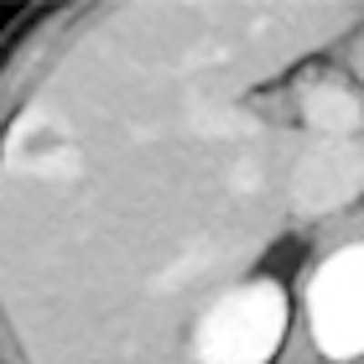
| Transverse abdominal CT images in a 51-year-old woman weighing 63 kg with a right hepatic lobe-enhancing lesion (hemangioma) reconstructed with FBP and three levels of ASIR (30%, 50%, and 70%) at four tube current-time products (200, 150, 100, and 50 mAs). Image noise and artifacts were reduced with ASIR, and images were diagnostically acceptable even at 50 mAs with 70% ASIR. Images republished with permission of the Radiological Society of North America from Radiology online before print, September 9, 2010, doi: 10.1148/radiol.10092212. |
There were no major artifacts seen in any of the ASIR or FBP image series acquired at the four dose levels; however, minor beam hardening or photon starvation artifacts were noted in both ASIR and FBP images in three of 22 patients.
Lesion conspicuity was significantly higher at 4.2 mGy on ASIR images compared to FBP images (observed p < 0.044), and overall diagnostic confidence also improved with the use of ASIR, changing from unacceptable on FBP to acceptable on ASIR images.
"Our study shows that reduction of radiation dose down to 8.4 mGy is possible when abdominal CT images are reconstructed with 30% ASIR blending and reduction of radiation dose down to 4.2 mGy is possible for patients weighing 90 kg or less with 50% and 70% ASIR blending," Singh and colleagues wrote.
Regardless of the dose levels, which ranged from 4.2 to 16.8 mGy, and ASIR percentage levels, subjective image noise was consistently reduced with ASIR versus FBP reconstructions. "This trend in subjective image noise was supported by a corresponding decrease in quantitative noise in ASIR images as opposed to FBP images," they added.
The results are in line with previous phantom studies, and with those of a smaller clinical study, the group noted. In that study (American Journal of Roentgenology, September 2009, Vol. 193:3, pp. 764-771), Hara and colleagues reported that ASIR can provide diagnostic quality images at 32% to 65% lower CTDIvol values than FBP techniques.
The authors cited the small sample size of the study and the presence of little variation in patient sizes as limitations. In addition, all scans were acquired in the equilibrium phase and the images were not examined during other phases, they wrote.
"Another implication of our study is the need to optimally set the correct ASIR-FBP blend," the authors noted. "For a CTDIvol of 8.4 mGy (x-ray tube current time-product of 100 mAs), 30% and 50% ASIR blending is appropriate; further dose reduction will require a higher degree of ASIR blending, with the associated image texture alterations."
Dose can be reduced further, to 4.2 mGy, by using 70% ASIR if radiologists are willing to accept a blotchy, pixilated image appearance, they added.
Reducing CT radiation dose to 8.4 mGy is feasible for abdominal CT images using ASIR reconstruction without compromising image quality, the group concluded. For patients weighing 90 kg or less, the dose may be reduced further to 4.2 mGy using ASIR.
"For routine abdominal CT examinations at 100 mAs in patients weighing greater than 90 kg, 30% and 50% ASIR-FBP blending provides acceptable image noise and diagnostic confidence, without a substantial change in image appearance," Singh and colleagues wrote.
By Eric Barnes
AuntMinnie.com staff writer
September 15, 2010
Related Reading
Better images are next frontier for CT iterative recon, August 30, 2010
ASIR halves VC dose without drop in image quality, June 21, 2010
MBIR aims to outshine ASIR for sharpness, CT dose reduction, May 18, 2010
Filtering software boosts CT image quality, lowers dose, May 7, 2010
ASIR cuts dose in Crohn's disease patients, December 4, 2009
Copyright © 2010 AuntMinnie.com


