Dual-source CT (DSCT) can reliably diagnose gout in its preclinical stages, opening the door to noninvasive diagnosis and earlier, more effective treatment, according to an article published in the April issue of the American Journal of Roentgenology.
Gout, a debilitating musculoskeletal disease that affects as many as 6 million people in the U.S., results from precipitation of monosodium urate crystals within joints. As described in a series published in the April issue of the American Journal of Roentgenology, Dr. Savvakis Nicolaou and colleagues from Vancouver General Hospital in British Columbia, Canada, took advantage of DSCT's ability to distinguish gout's monosodium urate crystal deposits from normal bone (AJR, April 2010, Vol. 194:4, pp. 1072-1078).
Difficult to diagnose
Gout can be difficult to diagnose using current methods, the authors explained.
"Although clinical features can help with the diagnosis of gout, many other diseases can mimic or coexist with it," wrote Nicolaou and colleagues including Charlotte Jane Yong-Hing and Dr. Sandro Galea-Soler. "Definitive diagnosis requires polarized light microscopy of fluid aspirated from the involved joint showing needle-shaped negatively birefringent monosodium urate crystals. Treatment is often initiated on assumption of the diagnosis. Joint aspiration can be technically difficult, and there is a risk of complications."
All of which makes a noninvasive diagnostic method highly desirable. Dual-source CT scanners (Somatom Definition, Siemens Healthcare, Malvern, PA) are equipped with two x-ray tubes, enabling simultaneous acquisition of images at two energy levels, creating two datasets that are loaded into a proprietary software package (syngo Dual Energy, Siemens) and read on the firm's CT workstation.
The typical scan protocol includes settings of 140 kV and 55 mAs for tube A, and 80 kV and 243 mAs for tube B, using a collimation of 0.6 mm and reconstructions of 0.75 mm. The total radiation dose is 2-3 mSv.
The proprietary software separates calcium from monosodium urate deposits using an image-based two-material decomposition algorithm that uses soft tissue as the baseline, the researchers explained. The attenuation differences in the two materials created by the high and low tube voltage acquisitions enable easy separation of monosodium urate (color-coded in red) from calcium (color-coded in blue).
 |
| syngo dual-energy CT software, Siemens Healthcare. The algorithm driving the syngo graphical user interface (above) plots uric acid versus calcium. The y-axis represents the attenuation values of the lower kilovoltage tube (80 kV), and the x-axis represents the attenuation values of the higher kilovoltage tube (140 kV). Pixels with a higher slope (high atomic number) are plotted above the line and represent calcium; the pixels below the line represent uric acid (lower atomic numbers of its component elements). Maximum and minimum values can be defined for soft tissue to improve performance of the algorithm. The algorithm ignores CT values outside this range. All images © 2010 American Roentgen Ray Society. |
Typically, the protocol includes bilateral imaging of several peripheral joints including elbows, wrists, hands, and feet.
Five patients
The paper describes five patients, including patient 1, a 54-year-old woman with a history of gout, who presented with painful dorsiflexion of the foot and a palpable mass along her posterior ankle. The radiologists aimed to determine whether the mass represented a tophus or an Achilles' heel rupture. Ultrasound showed enlargement of the tendon consistent with rupture. Dual-energy CT showed tendon enlargement but no uric acid, thus excluding gouty tophi as the cause of tendon rupture.
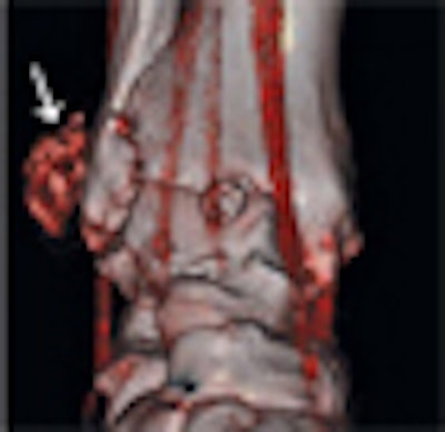
The second patient was a 45-year-old woman who presented to the emergency room with swelling along the right lateral malleolus but no history of trauma. Ultrasound depicted nonspecific soft-tissue swelling consistent with a mass or inflamed bursa, or potentially a soft-tissue sarcoma. Dual-energy CT confirmed that the mass was a tophus, a diagnosis confirmed by ultrasound-guided joint aspiration and mass aspiration.
 |
| A 45-year-old woman with swelling along lateral malleolus of right ankle but no history of trauma. Left, radiograph shows nonspecific soft-tissue swelling overlying lateral malleolus (arrow). Right, 3D volume-rendered coronal dual-energy CT image reveals that mass is composed of monosodium urate (red) consistent with tophus (arrow). |
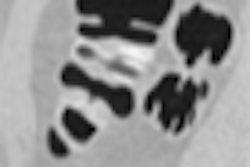
In the third case, a 50-year-old man presented to the emergency department after falling on his hand. Radiography revealed soft-tissue swelling but no fracture. Conventional CT showed abnormally high attenuation in the soft tissues along the carpus, but no fracture. Dual-energy CT revealed the presence of monosodium urate depositions.
Case 4 was of a 74-year-old man with chronic lymphocytic leukemia, who was admitted with a 24-hour history of increasing pain and swelling centered on the second toe proximal interphalangeal joint. Dual-energy CT showed the presence of a gouty tophus at the site of interest.
In case 5, a 71-year-old man presented to the emergency department with acute-onset pain in his left hand. Faint calcifications were visible on radiographs, while conventional CT showed areas of nodular thickening. Dual-energy CT revealed the presence of monosodium urate depositions along the joints.
Problem-solver
The five cases "show the utility of dual-energy CT as a problem-solving tool in identifying the presence of monosodium urate crystals and in confirming or excluding gout in clinically challenging cases," Nicolaou and colleagues wrote. "In cases 2, 3, and 5, the diagnosis of gout was confirmed by aspiration-proven monosodium urate deposition, and serum uric acid levels determined immediately after dual-energy CT were within the normal range. Markedly elevated serum uric acid levels were discovered in case 4, secondary to the underlying myeloproliferative disorder compounded with diuretic usage."
Dual-energy CT appears to be the only method of diagnosing such difficult cases with high accuracy, they wrote.
"The method's ability to detect the disease at its preclinical stage allows treatment aimed at preventing articular and bony damage to be started earlier, making disease regression easier to achieve and avoiding associated risks, such as increased cardiovascular mortality, decreased renal function, and formation of renal calculi," the authors wrote. "This benefit is important clinically because uric acid calculi respond well to medical therapy, thereby obviating more aggressive nonmedical treatments. In characterization of soft-tissue masses, dual-energy CT can accurately detect uric acid, thus obviating tissue aspiration, an invasive procedure."
By Eric Barnes
AuntMinnie.com staff writer
March 30, 2010
Related Reading
Dual-energy VC makes tagged materials disappear, October 29, 2008
DECT aces noninvasive gout diagnosis, September 15, 2008
Low-energy, high-current CT nabs tiny liver lesions, May 15, 2008
Osteoarthritis may predispose to acute gouty attacks, October 15, 2007
Fluoroscopically guided steroid injection effective in hip osteoarthritis, August 22, 2007
Copyright © 2010 AuntMinnie.com