Identifying fat within lesions is important for narrowing the range of differential diagnoses and potentially avoiding unnecessary intervention. For example, fatty lesions in the lungs and kidneys are usually benign.
CT and MRI are usually less effective for lesions with low fat concentrations, but would dual-energy CT be better at sussing out fat-containing pathologies? The picture is mixed, according to researchers in Israel.
Dual-energy CT images, whether from a dual-source scanner, a single-source dual-layer scanner, or a rapid-switching scanner, offer enhanced capacity for differentiation of tissues, a capability that remains underutilized clinically.
"The main advantages are better contrast and improved separation of high-density tissues," said radiologist Dr. Jacob Sosna from the Hadassah University Medical Center in Jerusalem.
In theory, fat has lower attenuation at low kVp than at high kVp, whereas water, bone, and iodine all have higher attenuation values at low kVp versus high kVp, Sosna said in a talk at the 2009 International Symposium on Multidetector-Row CT, sponsored by Stanford University of Stanford, CA. Therein lies dual-energy CT's theoretical ability to distinguish fatty from nonfatty tissues, a theory that has been shown to be helpful in a few clinical applications, but remains largely untested in others.
For its dual-energy imaging, the Jerusalem group uses an investigational scanner known as the Brilliance Simultaneous Dual Energy CT prototype (Philips Healthcare, Andover, MA). Dual-energy imaging is performed with a single x-ray tube, with photons aimed at two different scintillators in the detector made from different materials that are sandwiched together.
"Half of photons with lower energy are absorbed at the inner layer, and the outer layer absorbs the higher-energy photons and gave us the high-energy images," Sosna explained.
Some basic research was needed to evaluate dual-energy attenuation in different fatty materials, necessitating a two-part study that included a phantom and clinical components.
"In the phantom phase, the nature of changes in a phantom of different fat concentrations were assessed with dual-energy CT, and in the clinical phase, characterization of in vivo pathologies with different fat concentrations were also assessed," Sosna said.
The phantom was built from various materials from the grocery store, including butter (liquid, 82% fat), vegetable oil (60% fat), tahini (sesame paste, 60% fat), and water (0% fat). The materials were scanned using dual-energy protocols, and three images were created representing low-, high-, and combined-energy scans that mirror conventional MDCT.
Regions of interest were drawn on different areas of the test tubes for scans of different energy levels, Sosna said.
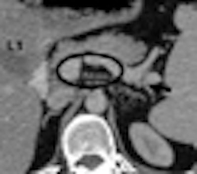
 |
| Measuring fatty-liver attenuation in regions of interest. All images and data courtesy of Dr. Jacob Sosna. |
Materials with the highest fat concentrations had appreciable HU differences between low and high kVp. The rest had far less difference.
"Only material with fat concentrations higher than 80% can lower the HU at the lower end of kVp," Sosna said. "So butter was -108 HU at lower energy and 105 HU at higher energy. However, intralipid [15% fat] yielded very similar numbers for the two energies: -13 for low kVp and -21 for high kVp."
 |
| When various materials were scanned at high-energy (L1 av HU) and low-energy (L0 av HU) (kVp) settings on a single-source dual-energy scanner, only materials with fat concentrations of > 80% showed significant differences in average attenuation between the two energy settings. |
The clinical arm of the study was a retrospective analysis of different fat-containing pathologies, using data from pathologies containing both macroscopic and microscopic fat. Regions of interest were drawn on the CT images for both low- and high-energy scans, and measurements were compared.
The group analyzed 33 patients (mean age, 56; range, 26-85) at 3-mm slice thickness, 1.5-mm reconstruction interval, 140 kVp, and 150 mAs.
Unlike with a dual-source scanner, using a single-source dual-detector system at 140 kVp means that half of the photons are low energy and half are high energy, Sosna explained. "The exact kVp depends on the patient's size and the body region scanned so that precise average energies can not be provided with this technique," Sosna told AuntMinnie.com.
The study included fatty livers, adrenal adenomas, and 19 other microscopic fat-containing lesions such as lipomas, renal angiomyolipomas, and adrenal myelolipomas, Sosna said. According to the results:
- Attenuation of fatty liver (n = 10) averaged 18 HU versus 25 HU at high energy.
- In four adrenal adenomas, HU was lower in low-energy images.
- For all other fatty findings (n = 19), HU was lower in low-energy images (-90 HU) compared to high-energy images (-78).
- The differences were statistically significant (p < 0.05) for each pathology type.
Although the high/low attenuation model worked well for some lesion types such as fatty liver, the differences were difficult to discern in others, such as adrenal adenomas, where only two cases followed the model. Some examples showed no discernable difference in attenuation.
 |
| Ten fatty livers scanned with dual-energy CT demonstrated significant differences between high-energy (HU L1) and low-energy (HU L0) scans; however, two of the four adrenal gland pathologies showed little difference in dual-energy scanning. |
 |
In 1991, Raptopoulos and colleagues first showed the value of dual-energy MDCT differentiating focal fatty infiltration from lower-density masses.
"The change in attenuation increased as the fat content in the liver increased," Raptopoulos et al wrote. "Analysis of the variance showed a statistically significant difference [p < 0.01] between fatty liver and other groups.... A difference greater than 10 HU was unique to fatty infiltration" (American Journal of Roentgenology, October 1991, Vol. 157:4, pp. 721-725).
Raptopoulos and colleagues "showed that different rates of HU were unique to fatty infiltration, which was also shown in our study," Sosna said.
The Jerusalem group's results are also in-line with those of Boland et al, who presented a study of difficult-to-discern adrenal gland pathology at the 2008 RSNA meeting, Sosna said.
In that dual-source CT study, "there was an average reduction of 3.3 HU between the 120-kV and 80-kV images; however, MDCT may not be helpful in characterizing lipid-poor adrenal adenomas," Sosna said of the group's conclusions.
Overall, Sosna's team found that "low-energy CT scans yielded lower CT values for lesions containing microscopic and macroscopic fat," he said. "Characterization is dependent on the fat concentration. There is need for assessment of detailed fat concentration sensitivity [of dual-energy CT] in comparison with MRI. Dual-energy will aid in the differentiation between fatty and solid lesions."
By Eric Barnes
AuntMinnie.com staff writer
July 8, 2009
Related Reading
MDCT holds promise for advanced osteoporosis scan, April 6, 2009
Rads prefer dual-energy x-ray over standard DR in lung, January 29, 2009
Adding CsI-based dual-energy imaging doesn't improve lung nodule detection, December 26, 2008
New CT method measures airway calcium to predict lung cancer, December 16, 2008
Dual-energy VC makes tagged materials disappear, October 29, 2008
Copyright © 2009 AuntMinnie.com


