
Advanced visualization analysis of MDCT images shows promise for improving the accuracy of imaging asbestos-related lung disease, according to physicians at the Barbara Ann Karmanos Cancer Institute in Detroit.
"By bringing the radiography of asbestos-related disease into the 21st century, we will reduce a lot of human suffering and save a lot of money," said Dr. Michael Harbut, co-director of the institute's National Center for Vermiculite and Asbestos-Related Cancers (NCVAC). Harbut discussed the use of 3D for asbestos imaging at the Asbestos Disease Awareness Organization (ADAO) conference last month in Manhattan Beach, CA.
Asbestos-related disease has traditionally been imaged using a plain posteroanterior and lateral chest film exam. These radiographs are read using the B-reader system, a network of physicians certified by the U.S. government for reviewing asbestos images. However, the method has been associated with high reader variability and is fraught with the potential for fraud in legal cases, Harbut said.
The Karmanos Cancer Institute typically uses 64-slice CT instead of performing chest radiography for asbestos-related disease. Led by NCVAC radiologist Dr. Carmen Endress, the institution has also begun utilizing advanced visualization software (Vitrea, Vital Images, Minnetonka, MN) on challenging cases. The software has provided valuable information in several applications, including identifying, tracking, and quantifying pulmonary fibrosis, he said.
 |
| Coronal CT image with segmented color fibrosis (left) and segmented thin-section color CT with fibrosis (right). All images courtesy of Dr. Michael Harbut of the Barbara Ann Karmanos Cancer Institute. |
Also, 3D software has proved handy in evaluating and monitoring pleural and pericardial plaques and in determining the cause of intractable pleural pain, he said.
"In asbestos-related disease, the most recent studies have shown that 40% of patients have some type of chest pain, which is likely noncardiac," he said. "A significant percentage of these patients go on to have intractable pleural pain that basically lasts forever and requires narcotics. Through the use of this technology, we think we've identified at least one of the mechanisms for intractable pleuratic pain."
 |
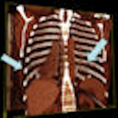
| Plaques on coronal oblique color-coded 3D CT image. |
 |
| Computed quantification of pleural plaque. |
The institute also believes that CT with advanced visualization software will help in identifying malignant lesions, although researchers do not yet have enough data to prove it, Harbut said.
Earlier diagnosis
In one recent case involving a worker at the Smithsonian Institution, the CT scan showed what appeared to be very early-stage asbestos-related disease.
"We [then] took his film and processed it through the [advanced visualization software] and found there to be more fibrotic change than we would expect," Harbut said. "It gave us one more piece of evidence in the diagnostic process to encourage me to tell people at the Smithsonian that they should really undertake screening of their remaining workers with similar exposure."
One downside to using 3D has been the time required to use the software; processing one patient can take anywhere from 20 minutes to an hour. However, the institute is working on automating aspects of that process, he said.
The technology might have its most significant contribution in reducing human suffering, Harbut said.
"I have a patient with intractable pleural pain who had a pleurectomy already, and we were trying to decide what to do with him because he has very advanced asbestosis," he said. "He's oxygen-dependent, he's got pleural plaques, and intractable pleural pain. We processed his film using the Vitrea system and found a pleural plaque rubbing on the internal aspect of the rib, creating a bony erosion."
 |
| Volume-rendered oblique coronal CT image with plaques leading to rib erosions. |
The software was able to obviate the need for another thoracoscopy and the use of narcotics. Instead, the patient was prescribed Marinol, and he was able to avoid the side effects of narcotics, he said.
"We also saved the system at least $15,000," Harbut said.
By Erik L. Ridley
AuntMinnie.com staff writer
April 16, 2009
Related Reading
Higher CT values associated with malignancy in lung nodules, April 14, 2008
Canadian CT lung screening tracks asbestos workers, April 3, 2008
Men show higher malignant mesothelioma rates after asbestos exposure, March 2, 2007
Indian shipbreakers suffering from asbestos -- report, September 9, 2006
Report links asbestos to larynx cancer, June 8, 2006
Copyright © 2009 AuntMinnie.com


