Optical colonoscopy could become safer, and polypectomies fewer, with the advent of a supermagnifying endoscope that can distinguish between benign hyperplastic lesions and potentially dangerous adenomas without removing them from the patient.
As many as half of the polyps removed at endoscopy are completely benign, so the new device could go a long way toward making colonoscopy safer, according to a gastroenterologist who presented an evaluation of the system at the 2009 Gastrointestinal Cancers Symposium in San Francisco.
Dr. Anna Buchner and colleagues from the Mayo Clinic in Jacksonville, FL, evaluated the ability of a probe-based confocal microscopic system (CFM) to help classify neoplastic and benign lesions in real-time during colonoscopy.
"The confocal probe-based microscopy system integrated into a dedicated endoscope shows excellent resolution of images during endoscopy, with the ability to achieve magnification of 1,000x, compared to zoom endoscopy, which gives us only 100x magnification, and normal endoscopy, which gives us only 30x magnification," Buchner said.
The recently developed CFM system uses an ultrathin probe (Minio probe, MKT, Orsay, France) that consists of thousands of fibers bundled together but is narrow enough (2.5 mm diameter) to be passed through the accessory channel of a standard endoscope, Buchner said. The confocal images are acquired during endoscopy at 12 frames per second and recorded before the scope is removed for later evaluation using software that captures and reconstructs the images, she said.
"The first aim of our study was to detect confocal image features of hyperplastic and neoplastic lesions," Buchner said. Researchers who were blinded and then unblinded to pathology results developed key features associated with neoplastic versus non-neoplastic lesions.
"The second aim was to measure the accuracy of confocal laser microscopy to distinguish neoplastic versus non-neoplastic colorectal lesions as compared to standard use of pathology," she said. "Our last aim was to assess the sensitivity, specificity, and positive and negative predictive value for classification of colorectal polyps detected using the new system."
The system was used in patients undergoing high-resolution colonoscopy. Once a lesion was detected, confocal images were obtained, followed by polypectomy and pathology, she said.
In this phase, confocal images of lesions were evaluated in a blinded fashion without the researchers' knowledge of their histologic diagnosis or endoscopic appearance. The confocal images were graded according to their crypt and nuclear characteristics based on features developed in the early phase of the study.
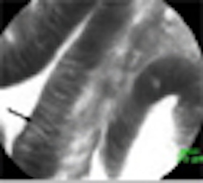
Confocal evaluation of villous adenomas revealed long, fingerlike "crypt" structures, while the adenocarcinomas in the series showed marked irregularities in the vasculatures, as well as in cell size and structure, she said.
The researchers assessed 81 polyps from 50 patients (mean age, 72). Of these 81 polyps, 30 were hyperplastic and 51 were neoplastic, with 39 tubular adenomas, seven tubulovillous adenomas, two villous, and three adenocarcinomas. The mean polyp size was 13 mm.
Standard histology found 30 hyperplastic polyps and 50 neoplastic polyps, whereas confocal imaging image interpretation showed 46 neoplastic and 35 hyperplastic polyps, Buchner said.
 |
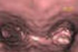
| (A) Endoscopic view of colon with confocal microscopic probe on polyp. (B) Endoscopic view alone. (C) Confocal endomicroscopy showing single cells and fingerlike villi typical of precancerous polyps. (D) Traditional view through a microscope after polypectomy shows the same villi as in image C. All images courtesy of Dr. Anna Buchner and the Mayo Clinic, Jacksonville, FL. |
From these data, the study group calculated that hyperplastic changes could be predicted with a sensitivity of 86%, a specificity of 93%, and an accuracy of 89%, with a positive predictive value of 95% and negative predictive value of 80%, she said.
"The confocal probe system allows accurate classification of colorectal lesions with malignant potential, and the use of this method has the potential to avoid polypectomies of non-neoplastic polyps," Buchner said.
A similar device is being developed for use in the upper digestive tract.
By Eric Barnes
AuntMinnie.com staff writer
January 30, 2009
Related Reading
Need for VC colon screening trumps physician turf battles, January 20, 2009
MR colonography separates benign from malignant colonic strictures, January 16, 2009
Study warns VC could leave high-risk polyps unresected, January 14, 2009
Slower not necessarily better in colonoscopy withdrawal, January 2, 2009
Colonoscopy cuts deaths from left-sided colon cancers only, December 16, 2008
Copyright © 2009 AuntMinnie.com