Researchers have recently looked into the willingness of zoos and veterinary schools to use their large-capacity CT scanners to image obese human patients. While the results showed that animal care facilities aren't a feasible solution for this need, a few facilities suggested they would consider a human exception.
Fortunately for four reptiles who recently underwent CT at a nonveterinary imaging center, some radiology practices also seem willing to consider the occasional exception to their usual patient group.
The four patients, all ailing Texas State Aquarium sea turtles, received much-needed assistance from group practice Radiology Associates of Corpus Christi, TX, at one of their outpatient imaging centers. CT showed that the sea turtles suffered from severe pneumonia and complications due to eating aquarium exhibits, among other conditions.
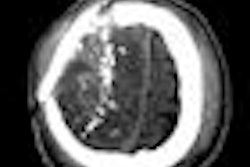
A CT scan of the pelvis and abdomen of Hemingway, a hawksbill sea turtle with an indiscriminate appetite, showed bolts from the aquarium's Tortuga Cay exhibit and an abscess in her intestines, according to a statement from the imaging provider.
"We actually saw bolts from the exhibit inside her," Texas State Aquarium dive officer and senior aquarist Deanna Gallier said. "She took a big bite out of the exhibit."
Hemingway and the other three turtles -- Itsy, Bitsy, and Daisy -- are from endangered species and are part of the Tortuga Cay sea turtle sanctuary at the Texas State Aquarium in Corpus Christi. All of the turtles were taken in as part of the aquarium's wildlife rehabilitation program, with injuries that deemed them unfit for release into the wild.
 |
| All images courtesy of Radiology Associates. |
Hemingway's troubles were signaled by an elevated white blood cell count and behavior changes, while Itsy and Bitsy presented with breathing problems and Daisy with a loss of appetite.
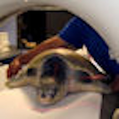
The four sea turtles received CT scans at the Radiology Associates Southside Imaging Center. Scans were performed on a Somatom Sensation 16 scanner (Siemens Healthcare, Malvern, PA), using a routine chest CT lung and soft-tissue protocol. A tube voltage of 120 kV and current of 400 mAs were used, with a 2-mm slice thickness and 2-mm reconstruction interval. Additional parameters included a gantry rotation time of 0.5 sec, detector collimation of 1.5 mm, and a 0.5 pitch.
 |
| CT scan of Hemingway's pelvis and abdomen revealed aquarium exhibit bolts and an abscess in her intestines. The bolts were successfully removed. |
The results showed that Itsy was suffering from severe pneumonia and Bitsy had a deviated lung, according to the statement. Daisy received happier news; instead of revealing serious illness or injury, her scan results showed that she was incubating several hundred eggs, potentially explaining her appetite loss.
Radiology Associates provides a wide range of services through its imaging centers, but its patient population is typically restricted to humans. It has, however, provided assistance to the aquarium in the past, helping a dolphin and Texas Indigo snake in need of services.
"One of our physicians was contacted through their personal relationship with one of the veterinarians that handles the aquarium," Radiology Associates administrator R. Ellis Keitt told AuntMinnie.com in an e-mail. "The aquarium approached us when they had an imaging need and we were happy that we could help them."
By Nicole Pettit
AuntMinnie.com staff writer
October 17, 2008
Related Reading
When zoos refuse: Obese patients face shortage of large-capacity, September 24, 2008
X-ray reveals python's too-hot dinner, July 21, 2006
X-ray alien head vanishes from duck stomach, May 26, 2006
Equine competitors get gold-medal care at Markopoulo clinic, August 26, 2004
Equine MRI clarifies navicular disease, August 22, 2004
Copyright © 2008 AuntMinnie.com