
As a method of imaging the endometrium, sonography has proved to be an excellent resource -- it doesn't impart radiation, it's low-cost, and it's fairly simple to use. And the endometrium's normal range of thickness has been well-defined with this modality.
But normal criteria for the thickness of the endometrium have not been established for CT, another modality commonly used for imaging the abdomen and pelvis. CTs performed for nongynecologic reasons sometimes reveal a thickened endometrium, which initiates a sonography referral.
However, often these results are not abnormal, according to researchers at Albert Einstein College of Medicine and Montefiore Medical Center in New York City. Dr. Julia Grossman and colleagues set out to study CT's ability to evaluate endometrial thickening, using transvaginal sonography as the reference standard, in the hope that establishing normal criteria can help women avoid unnecessary anxiety and facilities avoid unnecessary costs of additional imaging. They published their findings in this month's American Journal of Roentgenology (September 2008, Vol. 191:3, pp. 664-669).
"Our study shows that when CT depicts an apparently normal endometrial thickness, there is a high likelihood that the endometrium is indeed normal on sonography," the authors wrote.
The study included data gathered between March 2005 and January 2007 from 259 patients who had both transvaginal sonography and contrast-enhanced CT of the pelvis. Of the women who participated, 164 (63.3%) were premenopausal and 95 (36.7%) were postmenopausal.
Three readers (two board-certified radiologists and one abdominal imaging fellow) read sonograms and CT exams on different weeks; readers were blinded to identifying information and the results of the other technique. The readers knew patient age and last menstrual period or menopausal status when interpreting both exams.
Indications for CT included the following:
- Abdominal or pelvic pain
- Palpable abdominal masses
- Fever
- Hematuria
- Oncologic follow-up
- Nausea or bloating
- Ascites
- Follow-up for diverticulitis
CT scans were performed with either four-slice MDCT (LightSpeed Plus, GE Healthcare, Chalfont St. Giles, U.K.) or 64-slice MDCT (HiSpeed Advantage, GE). Sonography was performed on an Acuson XP or Sequoia system (Siemens Healthcare, Malvern, PA).
Grossman and colleagues assessed the endometrium with sonography by measuring it in the sagittal plane on its thickest portion. It was considered normal if its thickness was less than or equal to 16 mm in premenopausal women and less than or equal to 5 mm in postmenopausal women.
For CT, the endometrium was described as the hypoattenuated region and was evaluated by analyzing sequential axial images. The researchers classified the shape of the endometrium on CT as tubular or triangular, and they defined a normal endometrium as what would be expected for a patient's menstrual status and age; thickened was defined as greater than expected for the patient's menstrual status and age. A nonvisualized endometrium was categorized as one that was inseparable from the myometrium.
 |
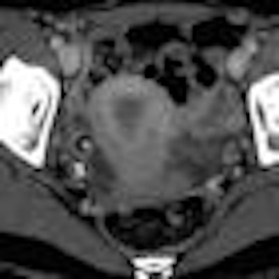
| Sixty-two-year-old woman with abnormal endometrium. Transvaginal sonogram (above) and axial contrast-enhanced CT image (below) show thickened endometrium in postmenopausal patient. Calipers (above) indicate thickness of endometrium. Images courtesy of the American Roentgen Ray Society; from Grossman J, Ricci Z, Rozenblit A, et al. Efficacy of contrast-enhanced CT in assessing the endometrium. AJR. 2008;191:664-669. |
 |
Of the 259 patients included in the study, CT identified the endometrium in 250. Of these 250 cases, there were 211 patients (84.4%) with normal and 39 patients (15.6%) with thickened endometrias.
Of the 250 patients whose endometrium was identified by CT, 201 had a normal and 49 had a thickened endometrium on sonography; of the 201 with a normal endometrium on sonography, the endometrium was visually normal on CT in 188 patients (93.5%), and of the 49 with a thickened endometrium on sonography, 26 (53.1%) also were thickened on CT. Results were falsely negative on CT in 23 of the 49 patients (47%) with thickened endometria.
 |
| Thirty-four-year-old woman with normal endometrium. Axial contrast-enhanced CT image (above) shows prominent triangular-shaped endometrium. Number 1 and circle indicate incidental measure of ovarian cyst not relevant to endometrium or this article. Sagittal reconstruction image (below) of same patient shows normal thin endometrium on this magnified image. Images courtesy of the American Roentgen Ray Society; from Grossman J, Ricci Z, Rozenblit A, et al. Efficacy of contrast-enhanced CT in assessing the endometrium. AJR. 2008;191:664-669. |
 |
Overall, Grossman's team found that the sensitivity and specificity of CT in detecting a thickened endometrium was 53.1% and 93.5%, respectively, compared to transvaginal sonography.
"Routine pelvic CT correctly identifies a normal endometrium in most patients," the researchers wrote. "CT is relatively insensitive in detecting the thickened endometrium but better able to identify gross rather than subtle thickening, which must be further characterized by transvaginal sonography."
By Kate Madden Yee
AuntMinnie.com staff writer
September 17, 2008
Related Reading
Study: MRI identifies submucosal fibroids that may migrate into endometrium, May 19, 2008
3D US of uterus a helpful adjunct to 2D scans, study finds, March 12, 2008
Copyright © 2008 AuntMinnie.com


