Multidetector CT offers many advantages over single-slice scanning, especially in noninvasive CT angiography. But artifacts can still mar MDCT images of the coronary arteries, especially if your scanner has only four detector rows.
Fortunately, most of these artifacts can be overcome with "judicious use of the appropriate image data reconstruction and display options," according to a new publication that summarizes common cardiac-scanning problems and their remedies.
Researchers from the Research Institute of Radiological Science and the Cardiovascular Center at Yonsei University College of Medicine in Seoul, South Korea, published their experience and images in the latest issue of RadioGraphics (May 2004, Vol. 24:3, pp. 788-800).
The group looked at 110 consecutive patients referred for CT angiography to exclude coronary artery disease or to evaluate coronary artery bypass grafts. Contrast-enhanced exams (1.25-mm collimation, 120 kV, 270-300 mA, 250-msec temporal resolution) were performed using a four-slice LightSpeed Plus (GE Healthcare, Waukesha, WI).
Cardiac images were routinely reconstructed from CT data acquired at 40% and at 70% of the R-R interval with a retrospective electrocardiographically gated algorithm. If those reconstructions didn't adequately depict the coronary arteries, subsequent tailored reconstructions were performed with data acquired at 30%, 50%, 60%, and 80%, the authors wrote.
The researchers then reviewed the axial source, as well as 3D volume-rendered, multiplanar reformatted, and maximum intensity projection images for artifacts, and classified the latter into four broad categories:
- Motion-related artifacts caused by cardiac, pulmonary, or other body motion.
- Beam-hardening effects caused by metallic implants, severe calcifications, or air bubbles in the pulmonary artery that obscured the underlying coronary vessel lumen.
- Structural artifacts produced by adjacent contrast material-filled structures and overlying vessels.
- Artifacts resulting from technical errors or limitations.
Altogether they identified 13 different types of problems affecting images from the 110 patients who were scanned. The group's publication includes a table describing the effects and suggested remedies, along with 18 figures showing problematic images.
Artifacts related to cardiac motion were the most common problem. In fact, despite the use of a retrospective ECG-gated reconstruction algorithm, images from all 110 patients were affected to some extent by factors such as higher heart rate, heart-rate variation during breathholds, arrhythmia, and inappropriate pitch selection.
 |
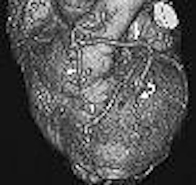
| Minimization of stepladder artifacts with maintenance of an optimal heart rate and selection of an optimal reconstruction window. Volume-rendered images reconstructed from data acquired at 40% (a, above) and 70% (b, below) of the R-R interval, with a heart rate of 55 beats per minute, show a stepladder artifact, which is less pronounced in (b). Note also the improved depiction in (b) of patency both in the in situ graft of the left internal mammary artery to the distal left anterior descending artery (arrowhead) and in the Y-graft (straight arrow) of a radial artery from the left internal mammary artery to the diagonal artery (curved arrow). Figure 5a, b, Choi HS, Choi BW, Choe KP, et al. Pitfalls, artifacts, and remedies in multi-detector row CT coronary angiography. RadioGraphics 2004; 23:787-800. |
 |
The cardiac motion artifacts generally occurred as blurring or stepladder effects; the easiest solution is the administration of beta blockers to lower the patient's heart rate.
"We found that a heart rate-lowering drug is routinely necessary to acquire diagnostic images, because a heart rate greater than 70-75 beats per minute, or variation in the heart rate during scanning, consistently induces artifacts," the authors wrote.
Reconstruction also plays a remedial role, the authors noted, as the various coronary arteries are best visualized at different points in the cardiac cycle.
"Reconstructed axial images, although apparently acquired in the same time window during the R-R interval, may differ in actual cardiac phase; the result of this disjuncture is an apparent stepladder-like contour of the heart," the authors wrote.
Overall, they noted, "Radiologists should be aware that an interpretation based exclusively on three-dimensional reconstructions of image data from the two separate phases of the cardiac cycle, without a review of axial source images, may be inaccurate and misleading."
"Every case with a suspected lesion should be interpreted by using all of the available reconstructions and with consideration for the anatomic relationships," the group concluded.
By Tracie L. ThompsonAuntMinnie.com staff writer
June 7, 2004
Related Reading
Sixteen-slice scanning extends CT's reach in coronary arteries, March 8, 2004
Task force recommends against routine coronary heart disease screening, February 17, 2004
CT coronary calcium attenuation affected by body mass, lesion location, January 28, 2004
Sixteen-slice CTA scanning makes everything better, January 5, 2004
EBCT versus MDCT? It depends on the application, July 01, 2003
Copyright © 2004 AuntMinnie.com